. 2010 Jan;18(1):19-34.
doi: 10.1007/s10577-009-9099-x.

Molecular analysis of the replication program in unicellular model organisms

Affiliations

Molecular analysis of the replication program in unicellular model organisms

M K Raghuraman et al. Chromosome Res. 2010 Jan.

Abstract

Eukaryotes have long been reported to show temporal programs of replication, different portions of the genome being replicated at different times in S phase, with the added possibility of developmentally regulated changes in this pattern depending on species and cell type. Unicellular model organisms, primarily the budding yeast Saccharomyces cerevisiae, have been central to our current understanding of the mechanisms underlying the regulation of replication origins and the temporal program of replication in particular. But what exactly is a temporal program of replication, and how might it arise? In this article, we explore this question, drawing again on the wealth of experimental information in unicellular model organisms.


Figures

Fig. 1
Fig. 1. The temporal program of replication illustrated by the kinetics of replication of four efficient origins in S. cerevisiae
Fig. 2
Fig. 2
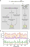
Whole-genome replication profiling. a Cartoon illustrating popular profiling methods. Density transfer experiments to separate replicated from unreplicated DNA or BrdU labeling to specifically label and isolate newly synthesized DNA can be used to construct replication profiles—progressive snapshots of the extent of replication across the genome—revealing general features of replication such as origin locations and the kinetics of replication for different genomic loci. Similar results can be obtained by copy number measurements. More precise determination of origin locations can be obtained by mapping ssDNA accumulated during S phase, occurring in the presence of HU. Mapping ssDNA in rad53 checkpoint mutants reveals all active origins, while similar experiments in wild-type cells reveals origins that fire predominantly early in S phase. Potential origins can be identified by chromatin immunoprecipitation (ChIP) of origin-binding proteins such as ORC followed by microarray hybridization or sequencing of the immunoprecipitated DNA. b Replication profiles for S. cerevisiae chromosome XI obtained by density transfer analysis at progressive S-phase times (pale blue through red profiles), ssDNA mapping (green profile), and ChIP of ORC and Mcm proteins (revealing potential origins; vertical orange bars). Yellow dot on x-axis, location of centromere. Profiles based on data from Feng et al. (2006), Xu et al. (2006), McCune et al. (2008)
Fig. 3
Fig. 3
The temporal program of replication. a Deterministic vs. stochastic models for origin firing time. In a strictly deterministic system, all cells in the population would fire their origins in the same temporal order (numbers above each bubble structure depicting the order of firing in that cell). In a strictly stochastic system, the firing sequence would be disordered. In a “controlled stochastic” or “fuzzy deterministic” system, chromatin or other forms of regulation could result in genomic regions showing differential access to critical initiation factors at different times in S phase, so that origins in different genomic zones could show different times of replication (red, yellow, and blue representing early-, mid-, and late-S zones; unfired pre-RCs indicated with red x marks in this simplistic view), but stochastic processes would still act locally, precluding a set order of origin activation. b One prediction of such a “controlled stochastic” model is that origins would show distributions of firing probabilities, with early and late origins showing distinct but potentially overlapping distributions (yellow), allowing for some disorder in the precise sequence of initiations but still resulting in a general temporal patterning of replication. c Firing probability distributions for S. cerevisiae ARS1413 on an early- (p13, red) vs. a late-replicating (pRightDup, blue) plasmid, inferred from the replication kinetic curves for these plasmids (Friedman et al. 1996; also see McCune et al. 2008 and Fig. 4). Because each plasmid had only one origin (ARS1413 in each case, with different amounts and arrangements of flanking genomic DNA included on the plasmid), the incremental change in percent replication (Δ% replication) reflects the incremental origin firing occurring per 2-min time interval through S phase
Fig. 4
Fig. 4
Cis-acting determinants of origin firing time in S. cerevisiae. Proximity to telomeres results in delayed firing of origins; chromatin is known to be a regulator of origin firing in this instance although the details of chromatin spreading from the telomere over 25+-kb distances are not well understood (Ferguson et al. 1991; Stevenson and Gottschling 1999; Cosgrove et al. 2002). Internal late-firing origins presumably are regulated by nontelomeric sequences proximal to the origins. One example is the cluster of late origins on S. cerevisiae chromosome XIV, where several sequences adjacent to the origins are known to act additively to delay origin firing time, as determined by monitoring the replication times of plasmids containing these origins along with different amounts of flanking sequence (Friedman et al. 1996). The precise nature of the timing determinants in this example is not known, but it has been shown that targeting a histone acetylase to one of the origins results in an advancement in its firing time (Vogelauer et al. 2002)
Nomenclature
Nomenclature
Origin efficiency: the percentage of cells in which an origin is observed to initiate DNA synthesis (or “fire”) in any given S phase. Origin competence: the percentage of cells in which an origin is biochemically competent to fire. Firing probability or instantaneous firing probability: the probability that an origin will fire during a particular interval within S phase. Firing rate or spatiotemporal initiation density: the number of initiations occurring per unit of time per length in the genome. The relationship between competence and origin efficiency is illustrated in the diagram above (modified from McCune et al. 2008). Depicted are three ARSs in a four-cell population as it enters S phase. An ARS has the potential to be used as an origin. It may be 100% competent if it successfully becomes biochemically competent to fire (red ring) in every cell in the population, or it may show <100% competence if it fails to achieve biochemical competence in some cells in the population. In those cells in which the origin failed to become competent, it would simply be replicated passively. The same fate may befall an origin that is competent but nevertheless is passively replicated by an incoming fork arriving at the origin before it has a chance to fire (broken red ring). Origin efficiency is the percentage of cells in which the origin actually fires

Similar articles

Cited by

References

    1. Alvino GM, Collingwood D, Murphy JM, et al. Replication in hydroxyurea: it's a matter of time. Mol Cell Biol. 2007;27:6396–6406. - PMC - PubMed
    1. Bell SP, Dutta A. DNA replication in eukaryotic cells. Annu Rev Biochem. 2002;71:333–374. - PubMed
    1. Bell SP, Stillman B. ATP-dependent recognition of eukaryotic origins of DNA replication by a multiprotein complex. Nature. 1992;357:128–134. - PubMed
    1. Blow JJ, Laskey RA. A role for the nuclear envelope in controlling DNA replication within the cell cycle. Nature. 1988;332:546–548. - PubMed
    1. Brewer BJ. When polymerases collide: replication and the transcriptional organization of the E. coli chromosome. Cell. 1988;53:679–686. - PubMed

Publication types

MeSH terms