

Mammographic density and matrix metalloproteinases in breast tissue
- PMID: 20012240
- PMCID: PMC2970805
- DOI: 10.1007/s12307-009-0031-x
Mammographic density and matrix metalloproteinases in breast tissue
Abstract
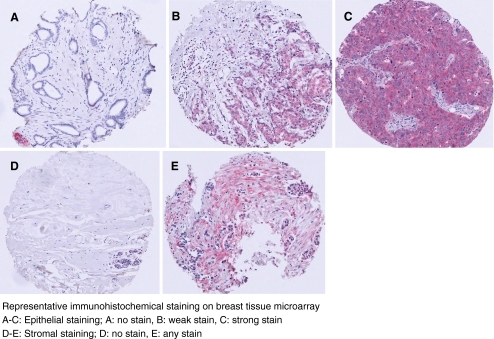
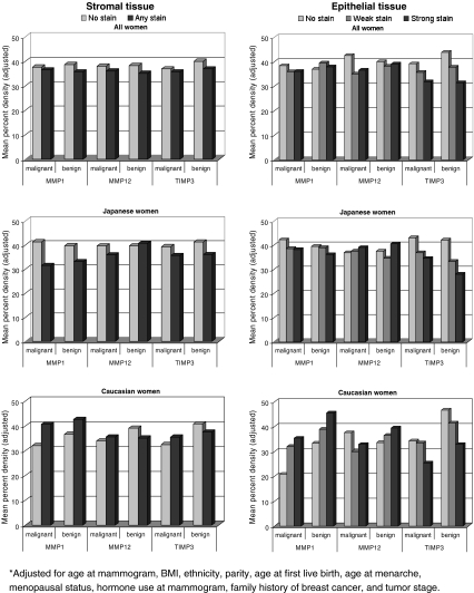
Mammographic density is a strong risk factor for breast cancer, yet the underlying histopathologic correlates are not clear. Matrix metalloproteinases (MMPs) and their inhibitors (TIMPs) play important roles in multiple stages of tumorigenesis. This study examined the association between mammographic density and expression of MMPs 1, 3, 9, and 12 and TIMP3 in benign and malignant breast tissue of 277 women with mainly Caucasian and Japanese ancestry. Tissue microarrays with up to 4 benign and 4 malignant cores per woman were stained immunohistochemically and evaluated. Digitized prediagnostic mammograms were assessed for densities using a computer-assisted method. General linear models adjusted for known confounders were applied to estimate mean densities by staining category. Strong expression of all MMPs was about twice as frequent in malignant as in benign tissue, while TIMP3 expression in stromal tissue was higher in benign than malignant cores. For MMP3 and 9, less than 10% of cores stained positive; thus, they were not further analyzed. None of the markers showed a statistically significant association with breast density in the entire study population and ethnic-specific results were conflicting and difficult to explain. Although not statistically significant, mean density was consistently lower with more extensive TIMP3 expression in stromal and epithelial tissue. These findings indicate that the higher breast cancer risk in women with dense breasts may be influenced by lower TIMP3 expression. However, future investigations into activities and ratios of additional proteases and their inhibitors as well as other pathways, such as inflammation, are needed.
Figures
Similar articles
-
Mammographic density and epithelial histopathologic markers.BMC Cancer. 2009 Jun 13;9:182. doi: 10.1186/1471-2407-9-182. BMC Cancer. 2009. PMID: 19523235 Free PMC article.
-
Strong association of tissue inhibitor of metalloproteinase (TIMP)-2 and -3 promoter single nucleotide polymorphisms with risk of colorectal cancer in ethnic Kashmiri population - a case control study.Biosci Rep. 2019 May 10;39(5):BSR20190478. doi: 10.1042/BSR20190478. Print 2019 May 31. Biosci Rep. 2019. PMID: 30988064 Free PMC article.
-
Matrix metalloproteinases MMP1, MMP2, MMP9 and their tissue inhibitors TIMP1, TIMP2, TIMP3 in head and neck cancer: an immunohistochemical study.Otolaryngol Pol. 2016 Jun 30;70(3):32-43. doi: 10.5604/00306657.1202546. Otolaryngol Pol. 2016. PMID: 27386931
-
Supplemental Screening for Breast Cancer in Women With Dense Breasts: A Systematic Review for the U.S. Preventive Service Task Force [Internet].Rockville (MD): Agency for Healthcare Research and Quality (US); 2016 Jan. Report No.: 14-05201-EF-3. Rockville (MD): Agency for Healthcare Research and Quality (US); 2016 Jan. Report No.: 14-05201-EF-3. PMID: 26866210 Free Books & Documents. Review.
-
An overview of mammographic density and its association with breast cancer.Breast Cancer. 2018 May;25(3):259-267. doi: 10.1007/s12282-018-0857-5. Epub 2018 Apr 12. Breast Cancer. 2018. PMID: 29651637 Free PMC article. Review.
Cited by
-
FKBP51 promotes invasion and migration by increasing the autophagic degradation of TIMP3 in clear cell renal cell carcinoma.Cell Death Dis. 2021 Oct 1;12(10):899. doi: 10.1038/s41419-021-04192-8. Cell Death Dis. 2021. PMID: 34599146 Free PMC article.
-
Plasma matrix metalloproteinases and postmenopausal breast cancer risk: a nested case-control study in the Multiethnic Cohort study.Breast Cancer Res Treat. 2012 Dec;136(3):837-45. doi: 10.1007/s10549-012-2308-x. Epub 2012 Oct 31. Breast Cancer Res Treat. 2012. PMID: 23112106 Free PMC article.
-
MMP-11 expression in early luminal breast cancer: associations with clinical, MRI, pathological characteristics, and disease-free survival.BMC Cancer. 2024 Mar 4;24(1):295. doi: 10.1186/s12885-024-11998-0. BMC Cancer. 2024. PMID: 38438841 Free PMC article.
-
Biological Mechanisms and Therapeutic Opportunities in Mammographic Density and Breast Cancer Risk.Cancers (Basel). 2021 Oct 27;13(21):5391. doi: 10.3390/cancers13215391. Cancers (Basel). 2021. PMID: 34771552 Free PMC article. Review.
-
Mammographic density as a predictor of breast cancer outcome.Future Oncol. 2010 Mar;6(3):351-4. doi: 10.2217/fon.10.3. Future Oncol. 2010. PMID: 20222792 Free PMC article.
References
Grants and funding
LinkOut - more resources
Full Text Sources
Miscellaneous