Review
. 2010 Jun;9(2):174-82.
doi: 10.1007/s12311-009-0147-z.

Computation of egomotion in the macaque cerebellar vermis

Affiliations
Review

Computation of egomotion in the macaque cerebellar vermis

Dora E Angelaki et al. Cerebellum. 2010 Jun.

Abstract

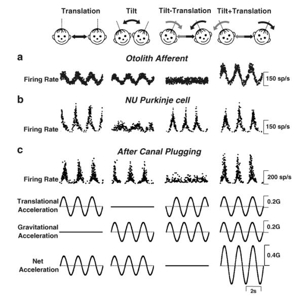
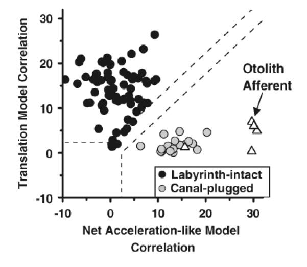
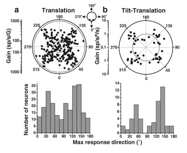
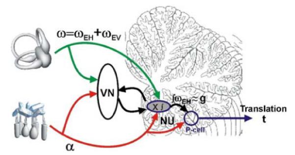
The nodulus and uvula (lobules X and IX of the vermis) receive mossy fibers from both vestibular afferents and vestibular nuclei neurons and are thought to play a role in spatial orientation. Their properties relate to a sensory ambiguity of the vestibular periphery: otolith afferents respond identically to translational (inertial) accelerations and changes in orientation relative to gravity. Based on theoretical and behavioral evidence, this sensory ambiguity is resolved using rotational cues from the semicircular canals. Recordings from the cerebellar cortex have identified a neural correlate of the brain's ability to resolve this ambiguity in the simple spike activities of nodulus/uvula Purkinje cells. This computation, which likely involves the cerebellar circuitry and its reciprocal connections with the vestibular nuclei, results from a remarkable convergence of spatially- and temporally-aligned otolith-driven and semicircular canal-driven signals. Such convergence requires a spatio-temporal transformation of head-centered canal-driven signals into an estimate of head reorientation relative to gravity. This signal must then be subtracted from the otolith-driven estimate of net acceleration to compute inertial motion. At present, Purkinje cells in the nodulus/uvula appear to encode the output of this computation. However, how the required spatio-temporal matching takes place within the cerebellar circuitry and what role complex spikes play in spatial orientation and disorientation remains unknown. In addition, the role of visual cues in driving and/or modifying simple and complex spike activity, a process potentially critical for long-term adaptation, constitutes another important direction for future studies.


Figures

Fig. 1
Fig. 1
Experimental protocol (top cartoons) and firing rates from (a) an otolith afferent, (b) simple spike responses of an NU Purkinje cell in labyrinthine-intact animals, and (c) simple spike responses of a NU Purkinje cell in canal-plugged animals. From left to right, stimuli were translation, tilt, tilt–translation, and tilt+translation (0.5 Hz), all delivered in complete darkness. Note that the translational and tilt stimuli were matched in both amplitude and direction to elicit an identical linear acceleration in the horizontal plane (bottom traces). Straight black and curved gray arrows denote translation and tilt axes of stimulation, respectively. Modified and replotted with permission from Angelaki [40] and Yakusheva [37]
Fig. 2
Fig. 2
Scatter plots of z-transformed partial correlation coefficients for the fits of each cell's responses with the “translation-coding” and “net acceleration-like” models. Black circles represent NU simple spike responses from labyrinthine-intact animals, whereas gray circles represent data from canal-plugged animals. Open triangles illustrate responses from otolith afferents. The superimposed dashed lines divide the plots into three regions: an upper/left area corresponding to cell responses that were significantly better fit (p<0.01) by the translation-coding model; a lower/right area that includes neurons which were significantly better fit by the net acceleration model; and an in-between area that would include cells that were not significantly better fit by either model. Modified and replotted with permission from Angelaki [40] and Yakusheva [37]
Fig. 3
Fig. 3
Preferred directions for simple spike responses to (a) translation and (b) tilt–translation (0.5 Hz). Top: Polar plot, where the radius corresponds to response gain and the polar angle illustrates the preferred (i.e., maximum response) direction in the horizontal plane. The inner and outer circles mark the tick marks of the gain scale. Each data point corresponds to one NU Purkinje cell. Bottom: Same data, now plotted as the distribution of preferred directions in the range (0°, 180°). Replotted with permission from Yakusheva [36]
Fig. 4
Fig. 4
Spatio-temporal matching of canal-driven (tilt−translation) and otolith-driven (translation) signals. (a) Distribution of the difference in preferred directions between the 0.5 Hz tilt−translation and translation stimulus conditions. (b, c) Response amplitude and phase during the 0.5 Hz tilt−translation stimulus (canal-driven component) is plotted as a function of the respective amplitude and phase during translation (otolith-driven component). Replotted with permission from Yakusheva [37]
Fig. 5
Fig. 5
Schematic illustrating our working hypothesis regarding the relationship between the processing of semicircular canal and otolith signals within the NU. Semicircular canal afferents carry head-referenced angular velocity (ω), but the NU encodes only the earth-horizontal component (ωEH). This signal is temporally integrated (ʃωEH) and used to cancel the gravitational component (g) of net linear acceleration (α), the signal carried by otolith afferents. The resulting output is the inertial component (t)

References

    1. Blazquez PM, Hirata Y, Highstein SM. The vestibulo-ocular reflex as a model system for motor learning: what is the role of the cerebellum? Cerebellum. 2004;3(3):188–192. - PubMed
    1. Boyden ES, Katoh A, Raymond JL. Cerebellum-dependent learning: the role of multiple plasticity mechanisms. Annu Rev Neurosci. 2004;27:581–609. - PubMed
    1. du Lac S, Raymond JL, Sejnowski TJ, Lisberger SG. Learning and memory in the vestibulo-ocular reflex. Annu Rev Neurosci. 1995;18:409–441. - PubMed
    1. Raymond JL, Lisberger SG, Mauk MD. The cerebellum: a neuronal learning machine? Science. 1996;272:1126–1131. - PubMed
    1. Barmack NH, Baughman RW, Errico P, Shojaku H. Vestibular primary afferent projection to the cerebellum of the rabbit. J Comp Neurol. 1993;327:521–534. - PubMed

Publication types

LinkOut - more resources