

Review
doi: 10.1007/978-3-642-02175-6_6.
Imaging of HIV/host protein interactions
Affiliations
- PMID: 20012526
- PMCID: PMC3209484
- DOI: 10.1007/978-3-642-02175-6_6
Item in Clipboard
Review
Imaging of HIV/host protein interactions
Curr Top Microbiol Immunol.
2009.
Abstract
HIV-1 relies on a myriad of interactions with host cell proteins to carry out its life cycle. Traditional biochemical approaches to probe protein-protein interactions are limited in their ability to study the spatial and dynamic interactions that take place in the context of an intact cell. However, issues such as localization and dynamics of interactions between viral and host proteins can be well addressed utilizing fluorescent imaging methods. The past decade has brought about the development of many novel fluorescent imaging techniques which have proved useful to describe the interaction of HIV-1 proteins with the host cell.
Figures
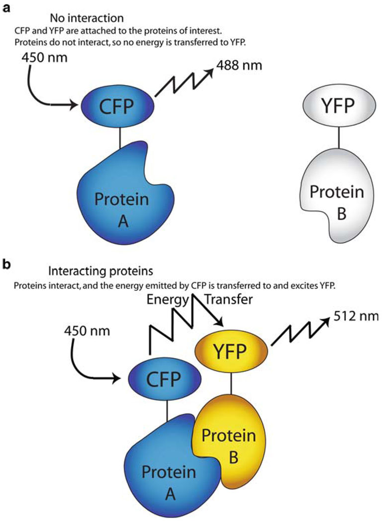
Fluorescence resonance energy transfer (FRET). (a) No interaction. CFP and YFP are attached to the proteins of interest. Proteins do not interact, so no energy is transferred to YFP. (b) Interacting proteins. Proteins interact, and the energy emitted by CFP is transferred to and excites YFP

Bifluorescence complementation (BiFc). (a) No interaction. GFP is split into two halves which cannot fluoresce alone and attached to the proteins of interest. Proteins do not interact, so no fluorescent signal is produced. (b) Interacting proteins. Proteins interact, allowing the two halves of GFP to come together and produce a fluorescent signal

Total internal reflection fluorescence (TIRF) microscopy. Approaching light at the critical angle undergoes total internal reflection back through the glass coverslip An evanescent wave is created on the other side of the glass, extending approximately 100 nm into the sample. As only this extremely thin region is illuminated, background fluorescence from out-of-focus light is dramatically decreased, creating greater sensitivity

HIV-1 Life Cycle

GFP-Vpr can be used to visualize HIV-1 trafficking within living cells
Similar articles
-
Application of Advanced Light Microscopy to the Study of HIV and Its Interactions with the Host.Viruses. 2021 Feb 1;13(2):223. doi: 10.3390/v13020223. Viruses. 2021. PMID: 33535486 Free PMC article. Review.
-
Investigation of HIV-1 assembly and release using modern fluorescence imaging techniques.Traffic. 2013 Jan;14(1):15-24. doi: 10.1111/tra.12006. Epub 2012 Oct 3. Traffic. 2013. PMID: 22957540 Review.
-
Imaging Viral Infection by Fluorescence Microscopy: Focus on HIV-1 Early Stage.Viruses. 2021 Jan 30;13(2):213. doi: 10.3390/v13020213. Viruses. 2021. PMID: 33573241 Free PMC article. Review.
-
Where in the Cell Are You? Probing HIV-1 Host Interactions through Advanced Imaging Techniques.Viruses. 2016 Oct 19;8(10):288. doi: 10.3390/v8100288. Viruses. 2016. PMID: 27775563 Free PMC article. Review.
-
Live cell visualization of the interactions between HIV-1 Gag and the cellular RNA-binding protein Staufen1.Retrovirology. 2010 May 10;7:41. doi: 10.1186/1742-4690-7-41. Retrovirology. 2010. PMID: 20459747 Free PMC article.
Cited by
-
A SNAP-tagged derivative of HIV-1--a versatile tool to study virus-cell interactions.PLoS One. 2011;6(7):e22007. doi: 10.1371/journal.pone.0022007. Epub 2011 Jul 22. PLoS One. 2011. PMID: 21799764 Free PMC article.
-
Help or Hinder: Protein Host Factors That Impact HIV-1 Replication.Viruses. 2024 Aug 10;16(8):1281. doi: 10.3390/v16081281. Viruses. 2024. PMID: 39205255 Free PMC article. Review.
-
A model for cofactor use during HIV-1 reverse transcription and nuclear entry.Curr Opin Virol. 2014 Feb;4(100):32-6. doi: 10.1016/j.coviro.2013.11.003. Epub 2014 Jan 14. Curr Opin Virol. 2014. PMID: 24525292 Free PMC article. Review.
References
-
- Ando R, Mizuno H, Miyawaki A. Regulated fast nucleocytoplasmic shuttling observed by reversible protein highlighting. Science. 2004;306:1370–1373. - PubMed
-
- Arhel N, Genovesio A, Kim K, Miko S, Perret E, Olivo-Marin J, Shorte S, Charneau P. Quantitative four-dimensional tracking of cytoplasmic and nuclear HIV-1 complexes. Nat Methods. 2006;3:817–824. - PubMed
-
- Axelrod D, Burghardt TP, Thompson NL. Total internal reflection fluorescence. Annu Rev Biophys Bioeng. 1984;13:247–268. - PubMed
-
- Bailey B, Farkas DL, Taylor DL, Lanni F. Enhancement of axial resolution in fluorescence microscopy by standing-wave excitation. Nature. 1993;366:44–48. - PubMed
Publication types
MeSH terms
Grants and funding
LinkOut - more resources
Full Text Sources
Miscellaneous
