

Review
doi: 10.1007/978-3-642-02175-6_7.
Virological and cellular roles of the transcriptional coactivator LEDGF/p75
Affiliations
- PMID: 20012527
- PMCID: PMC3093762
- DOI: 10.1007/978-3-642-02175-6_7
Item in Clipboard
Review
Virological and cellular roles of the transcriptional coactivator LEDGF/p75
Curr Top Microbiol Immunol.
2009.
Abstract
The chromatin-associated cellular proteins LEDGF/p75 and LEDGF/p52 have been implicated in transcriptional regulation, cell survival and autoimmunity. LEDGF/p75 also appears to act as a chromatin-docking factor or receptor for HIV-1 and other lentiviruses and to play a role in leukemogenesis. For both the viral and cellular roles of this protein, a key feature is its ability to act as a molecular adaptor and tether proteins to the chromatin fiber. This chapter reviews the emerging roles of LEDGF/p75 and LEDGF/p52 in diverse cellular processes and disease states.
Figures
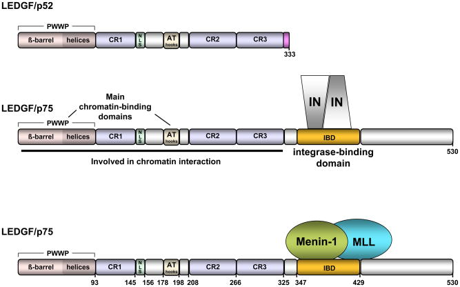
Domain structure and binding partners of LEDGF/p75 and LEDGF/p52.
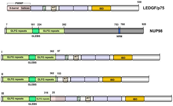
NUP98 contains eight N- terminal GLFG repeats that govern localization of the protein to nuclear GLFG bodies. GLEBS is a sequence within the GLFG repeats that serves as docking site to GLE2p, the yeast ortholog of human RAE1 a protein involved in mRNA nuclear export. Nucleoporin RNA binding motif (NRM) is an octapeptide with partial homology to the ribonucleoprotein motif. In the NUP98-LEDGF/p75 fusion proteins (I–III) described in leukemic patients, the NUP98 GLFG repeats and the GLEBS element fuse to LEDGF/p75 segments that contain the IBD.
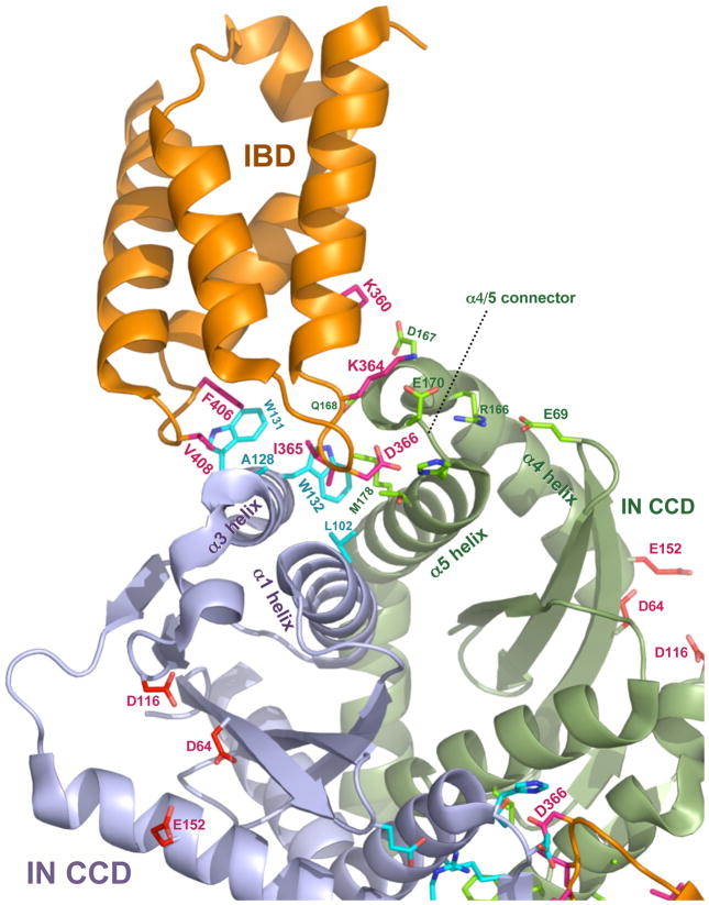
The figure was constructed with MacPyMOL from Protein Data Bank file 2BJ4 (www.pdb.org , ref. [13]. LEDGF/p75 contributes most of the amino acid side chains that make direct contact with IN. D366 engages in a pair of essential hydrogen bonds with the main chain amides of E170 and H171 in the alpha-4/5 connector of one IN monomer. Hydrophobic interactions predominate in interactions with the other IN monomer: IN residues W132, W131, A128, L102 form a pocket in the vicinity of IBD residues F406 and V408; this pocket buries IBD residue I365). IN catalytic center resides D64, D116 and E152, the mutation of which produce purely catalytic defects (reviewed in [25]) are shown in the lower half of the figure for each IN monomer. Interaction between the HIV-1 IN NTD and the IBD was previously established by biochemical evidence [53] and Hare et al. have recently solved a co-crystal structure for the LEDGF/p75 IBD complexed with a two-domain fragment of HIV-2 IN (NTD+CCD) [38]. Extensive structural contacts between the IBD and the NTD were identified and characterized [38]. Charged interactions are dominant, with conserved acidic residues in the HIV-2 IN NTD (E6, E10, E13) engaging complementary basic residues in the IBD (K401, R404, R405). Moreover, the NTDs of other lentiviral INs contain the same or closely adjacent glutamic acid residues. Enhancement by LEDGF/p75 of concerted strand transfer activity in vitro was also shown to be impaired by charge-reversing mutation of the basic IBD residues to glutamates in the equine lentivirus (EIAV) IN [38]. This activity could then be partially restored by reciprocal mutation of the acidic IN residues to lysines, verifying that these charged interactions are the main structural feature. See ref. [17] for a recent review of HIV-1 IN structural biology.
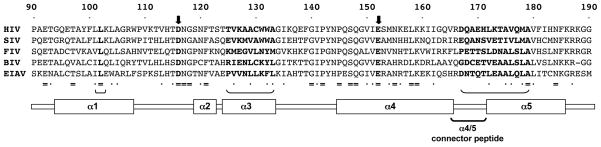
Alignment of the central part of the CCD for IN proteins from the three lentiviral subgenera (primate, feline, ungulate). Identity is indicated by = and residues with conserved biochemical features by dots. IN alpha helices are indicated below and the segments primarily involved in forming the IBD binding pocket are indicated by bold-face font and brackets. The relative lack of sequence conservation in these regions, e.g., the alpha 4/5 connector, is evident. Figure 2 shows the placement of these protein elements for the interface of HIV-1 IN with LEDGF/p75; catalytic center residues (heavy black arrows here) do not contact the IBD. Although its exact oligomerization state in the PIC is not conclusively established, the weight of evidence is in favor of the enzyme acting as a multimer, with a tetramer likely [4, 15, 30, 31, 37, 39, 40, 46, 84, 96]. In vitro, an IN dimer enables 3′ end processing but a tetramer appears needed for DNA strand transfer activity [30, 37, 46]. Of note, higher order multimers were defined in the Hare et al. co- crystal structure of the IBD with HIV-2 INNTD+CCD [38]. Their relevance to the oligmeric state in the virus remains to be determined.
Similar articles
-
Alternative splicing and caspase-mediated cleavage generate antagonistic variants of the stress oncoprotein LEDGF/p75.Mol Cancer Res. 2008 Aug;6(8):1293-307. doi: 10.1158/1541-7786.MCR-08-0125. Mol Cancer Res. 2008. PMID: 18708362 Free PMC article.
-
LEDGF/p75 TATA-less promoter is driven by the transcription factor Sp1.J Mol Biol. 2011 Nov 25;414(2):177-93. doi: 10.1016/j.jmb.2011.10.010. Epub 2011 Oct 12. J Mol Biol. 2011. PMID: 22019592
-
Characterization of the HIV-1 integrase chromatin- and LEDGF/p75-binding abilities by mutagenic analysis within the catalytic core domain of integrase.Virol J. 2010 Mar 23;7:68. doi: 10.1186/1743-422X-7-68. Virol J. 2010. PMID: 20331877 Free PMC article.
-
The lentiviral integrase binding protein LEDGF/p75 and HIV-1 replication.PLoS Pathog. 2008 Mar 28;4(3):e1000046. doi: 10.1371/journal.ppat.1000046. PLoS Pathog. 2008. PMID: 18369482 Free PMC article. Review.
-
Integrase, LEDGF/p75 and HIV replication.Cell Mol Life Sci. 2008 May;65(9):1403-24. doi: 10.1007/s00018-008-7540-5. Cell Mol Life Sci. 2008. PMID: 18264802 Free PMC article. Review.
Cited by
-
Absence of LEDGF/p75 Expression in Astrocytes May Affect HIV-1 Integration Efficiency.Mol Gen Microbiol Virol. 2019;34(2):81-83. doi: 10.3103/s0891416819020113. Epub 2019 Oct 14. Mol Gen Microbiol Virol. 2019. PMID: 33867663 Free PMC article.
-
The same pocket in menin binds both MLL and JUND but has opposite effects on transcription.Nature. 2012 Feb 12;482(7386):542-6. doi: 10.1038/nature10806. Nature. 2012. PMID: 22327296 Free PMC article.
-
Integrating the HIV-1 assembly/maturation pathway.Proc Natl Acad Sci U S A. 2013 May 21;110(21):8327-8. doi: 10.1073/pnas.1306620110. Epub 2013 May 13. Proc Natl Acad Sci U S A. 2013. PMID: 23671082 Free PMC article. No abstract available.
-
A role for microRNA-155 modulation in the anti-HIV-1 effects of Toll-like receptor 3 stimulation in macrophages.PLoS Pathog. 2012 Sep;8(9):e1002937. doi: 10.1371/journal.ppat.1002937. Epub 2012 Sep 20. PLoS Pathog. 2012. PMID: 23028330 Free PMC article.
-
Viral DNA tethering domains complement replication-defective mutations in the p12 protein of MuLV Gag.Proc Natl Acad Sci U S A. 2013 Jun 4;110(23):9487-92. doi: 10.1073/pnas.1221736110. Epub 2013 May 9. Proc Natl Acad Sci U S A. 2013. PMID: 23661057 Free PMC article.
References
-
- Ahuja HG, Hong J, Aplan PD, Tcheurekdjian L, Forman SJ, Slovak ML. t(9;11)(p22;p15) in acute myeloid leukemia results in a fusion between NUP98 and the gene encoding transcriptional coactivators p52 and p75-lens epithelium-derived growth factor (LEDGF) Cancer Res. 2000;60(22):6227–9. - PubMed
-
- Ahuja P, Caffe AR, Holmqvist I, Soderpalm AK, Singh DP, Shinohara T, van Veen T. Lens epithelium-derived growth factor (LEDGF) delays photoreceptor degeneration in explants of rd/rd mouse retina. Neuroreport. 2001;12(13):2951–5. - PubMed
-
- Argiropoulos B, Humphries RK. Hox genes in hematopoiesis and leukemogenesis. Oncogene. 2007;26(47):6766–76. - PubMed
-
- Bao KK, Wang H, Miller JK, Erie DA, Skalka AM, Wong I. Functional oligomeric state of avian sarcoma virus integrase. J Biol Chem. 2003;278(2):1323–7. - PubMed
-
- Bartholomeeusen K, De Rijck J, Busschots K, Desender L, Gijsbers R, Emiliani S, Benarous R, Debyser Z, Christ F. Differential Interaction of HIV-1 Integrase and JPO2 with the C Terminus of LEDGF/p75. J Mol Biol. 2007;372(2):407–21. - PubMed
Publication types
MeSH terms
Substances
Grants and funding
LinkOut - more resources
Full Text Sources
Research Materials
