Review
. 2010 Feb;67(4):545-56.
doi: 10.1007/s00018-009-0209-x. Epub 2009 Dec 12.

Platelets and viruses: an ambivalent relationship

Affiliations
Review

Platelets and viruses: an ambivalent relationship

Claire Flaujac et al. Cell Mol Life Sci. 2010 Feb.

Abstract

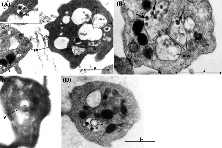
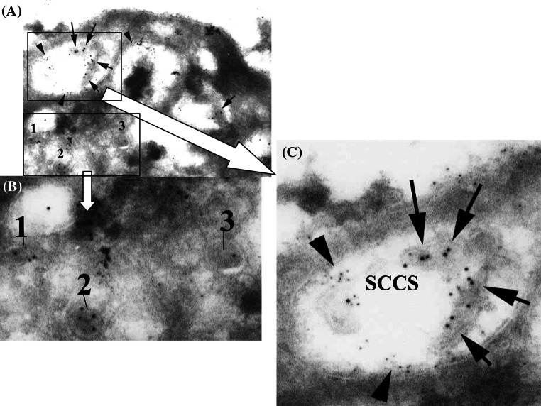
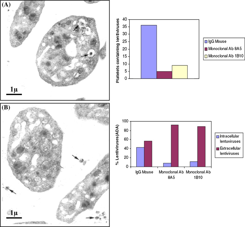
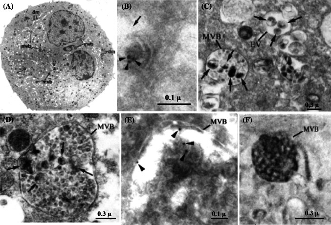
Thrombocytopenia is a frequent complication of viral infections providing evidence that interaction of platelets with viruses is an important pathophysiological phenomenon. Multiple mechanisms are involved depending on the nature of the viruses involved. These include immunological platelet destruction, inappropriate platelet activation and consumption, and impaired megakaryopoiesis. Viruses bind platelets through specific receptors and identified ligands, which lead to mutual alterations of both the platelet host and the viral aggressor. We have shown that HIV-1 viruses are internalized specifically in platelets and megakaryocytes, where they can be either sheltered, unaltered (with potential transfer of the viruses into target organs), or come in contact with platelet secretory products leading to virus destruction and facilitated platelet clearance. In this issue, we have reviewed the various pathways that platelets use in order to interact with viruses, HIV and others. This review also shows that more work is still needed to precisely identify platelet roles in viral infections, and to answer the challenge of viral safety in platelet transfusion.


Figures

Fig. 1
Fig. 1
EM examination of platelets incubated with HIV viruses. Characteristic images of intact HIV viruses are observed: a in small endocytic vesicles (VI) and b,d in the open (surface connected) canalicular system (OCS). The viruses display round shape, dense core and intact envelope. c P24 immunolabeling confirm that HIV viruses are found in endocytic vesicles (V)
Fig. 2
Fig. 2
a Platelets incubated with VSV lentiviruses, having undergone double immunolabeling for the viral protein P24(10 nm gold, arrows) and the α-granule marker P-selectin (5 nm gold, arrowheads). b Three lentiviruses (1, 2, 3), identified by P24 immunolabeling, can be detected in small endocytic vesicles deprived of α-granule marker. These viruses display round shape and intact envelope. c Double immunolabeling for the viral protein P24 and the α-granule marker P-selectin (b). On the other hand, lentiviruses present in the surface connected canalicular system (SCCS) can only be identified by P24 immunolabeling (arrows). Indeed, they are degraded, having lost their viral envelope and their round morphology. The presence of P-selectin on the surface connected canalicular system (SCCS) membrane (arrowheads) attests for granular secretion. This indicates that contact with platelet secretory products is able to damage internalized viruses (bar 0.125 μm)
Fig. 3
Fig. 3
Diagram of the various platelet receptors involved in platelet–virus interaction
Fig. 4
Fig. 4
The two pathways of HIV internalization within host cells
Fig. 5
Fig. 5
Demonstration of DC-SIGN expression on platelets: a Detection of DC-SIGN expression by flow cytometry on platelets and dendritic cells. Platelets: DC-SIGN labeling shows that 15% of platelets are DC-SIGN positive. Dendritic cells: as a positive control, an equivalent proportion of dendritic cells express DC-SIGN by flow cytometry. b Immunogold labeling for DC-SIGN: DC-SIGN antigen (arrows) is detected on platelet membrane, on SCCS membrane, and on some limiting membrane α-granules (bar 0.3 lm). c DC-SIGN detection in platelets by western blot. DC-SIGN, a 44-kDa protein, is detected in platelets, and in Hela-DC-SIGN and THP1-DC-SIGN cells (positive control). Hela and THP-1 cells are negative controls
Fig. 6
Fig. 6
DC-SIGN in platelets is functional. a Platelet samples incubated with HIV-e lentiviruses in the absence of anti-DC-SIGN antibody: virtually no extra-cellular viruses are found, whereas numerous images of intracellular viruses can be seen (arrows) (A = α-granules). b Platelet sample incubated with HIV-e lentiviruses in the presence of anti-DC-SIGN antibody: most viruses were observed outside platelets (arrows), and virtually none inside them. Graph Comparison of the respective proportions of extracellular and intracellular lentiviruses as observed on the control samples and in the presence of anti-DC SIGN antibody
Fig. 7
Fig. 7
a EM view of a characteristic mature cultured MK showing demarcation membranes (dm), alpha granules (a), multivesicular bodies (mvb), and incubated with VSV-e lentivirus suspension: b Lentiviruses located in endocytic vacuoles are identified thanks to their size and specific P24 immunolabeling (arrowheads). c They are also found (arrows) in small endocytic vesicles and multivesicular bodies (MVB). In the EV (arrow), viruses seem intact, displaying spherical shape and well-limited envelope. d When located in the MVB, lentiviruses (arrows) appear swollen and poorly limited, with irregular shape. e P24 immunolabeling is detected in MVB (arrowheads), confirming the presence of lentiviruses in these structures. fMVBs are positive for acid phosphatase (revealed by the cerium cytochemical technique) which contributes to viral destruction
Fig. 8
Fig. 8
EM view of a macrophage from a mouse infected by adenoviruses: numerous platelets (P) adhere to the macrophage surface, one of them being partly degranulated displays an intracellular virus-like particle (red arrow). Inset EM aspect of the adenovirus preparation

References

    1. Danon D, Jerushalmy Z, De Vries A. Incorporation of influenza virus in human blood platelets in vitro electron microscopical observation. Virology. 1959;9:719–722. doi: 10.1016/0042-6822(59)90168-0. - DOI - PubMed
    1. Jerushalmy Z, Kohn A, De Vries A. Interaction of myxoviruses with human blood platelets in vitro. Proc Soc Exp Biol Med. 1961;106:462–466. - PubMed
    1. White JG, Clawson CC. Effects of small latex particle uptake on the surface connected canalicular system of blood platelets: a freeze-fracture and cytochemical study. Diagn Histopathol. 1982;5:3–10. - PubMed
    1. Youssefian T, Drouin A, Masse JM, Guichard J, Cramer EM. Host defense role of platelets: engulfment of HIV and Staphylococcus aureus occurs in a specific subcellular compartment and is enhanced by platelet activation. Blood. 2002;99:4021–4029. doi: 10.1182/blood-2001-12-0191. - DOI - PubMed
    1. Pelletier JP, Transue S, Snyder EL. Pathogen inactivation techniques. Best Pract Res Clin Haematol. 2006;19:205–242. doi: 10.1016/j.beha.2005.04.001. - DOI - PMC - PubMed

MeSH terms

Substances