. 2010 Feb;34(2):271-81.
doi: 10.1007/s00264-009-0924-9. Epub 2009 Dec 12.

Nonunions around the knee joint

Affiliations

Nonunions around the knee joint

Daniel B Chan et al. Int Orthop. 2010 Feb.

Abstract

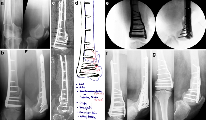
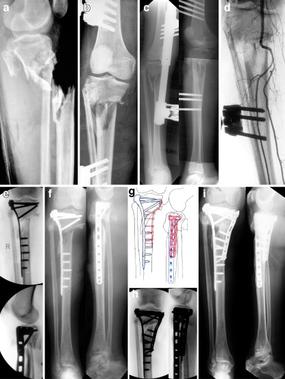
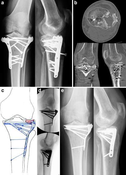
Nonunions of the distal femur are relatively rare but can occur with or without previous surgery. Many surgical methods have been described for the treatment of these nonunions including external fixation, intramedullary nailing, arthroplasty, and fixed angle plating. While arthroplasty may have a limited role in the elderly patient with osteoporosis and severe communiution, most nonunions can be reliably treated with revision fixation using a fixed angled device, lag screws, and bone grafting as needed. Fixed-angle plating also has the advantage of stabilising a small distal segment where intramedullary nailing has proven to be less effective. Any surgical intervention must include restoration of the normal mechanical axis of the distal femur for a satisfactory clinical outcome. Proximal tibia nonunions are a similarly rare clinical entity due to the excellent blood supply in the metaphysis. Extra-articular proximal tibial nonunions are often due to malreduction during the index surgery resulting in poor bone contact and biomechanical stability. Soft tissue forces and improper use of intramedullary implants often predispose these fractures to malalignment. Revision surgery consisting of deformity correction and at least a lateral tension band plate with lag screws has led to predictable union and good clinical results. Tibial plateau nonunions are exceedingly uncommon but are difficult to treat because of small, often devascularised fragments, post-surgical scarring, and osteopenia. Revision surgery with meticulous soft tissue handling to restore the articular surface and mechanical axis with repair of meniscal pathology gives the patient the best chance of recovering knee function.


Figures

Fig. 1
Fig. 1
A 50-year-old female was involved in a motor vehicle accident and sustained a right-sided supracondylar distal femur fracture. Locking plate fixation was placed percutaneously using a less invasive stabilization system (LISS) plate and screws. She was diagnosed with a nonunion at six months and surgical revision was planned and performed with debridement and osteotomy of the nonunion site, placement of demineralized bone matrix (DBM) bone graft, and a medially placed locking compression plate (LCP) and screws including interfragmentary lag screws and screws for additional compression and stability. She returned at regular follow-up intervals and healed uneventfully. The LISS plate was removed two years following revision surgery due to tenderness near the plate. She last returned at 39 months following surgical revision with excellent clinical and radiographic results including a healed distal femur fracture, resolution of pain symptoms, and a return to all pre-injury activities. a Anteroposterior (AP) and lateral injury radiographs illustrating a right-sided supracondylar distal femur fracture. b AP and lateral films at six months revealing a nonunion. c CT scan images further illustrating the nonunion. d Preoperative plan for revision surgery. e Intraoperative fluoroscopic AP and lateral images. f AP and lateral images at one year following revision surgery revealing a healed distal femur nonunion. g AP and lateral images at 39 months (and 18 months following removal of the lateral plate and screws)
Fig. 2
Fig. 2
A 65-year-old female was skiing and was struck by another skier who was airborne after going off of a jump. She sustained a right-sided Grade IIIC open proximal tibia fracture with segmental metaphyseal bone loss. She was taken to a local trauma centre and also had extensive degloving. A joint spanning external fixator was applied and a vascular reconstruction was performed bypassing her popliteal trunk. She was transferred to our institution at three months following injury for definitive management of her tibia fracture. Presence of an eschar was noted near the fracture site and she had a viable foot but did have a foot drop with numbness on the dorsum and distal plantar aspect of her foot. She underwent multiple irrigation and debridements followed by removal of the external fixator, open reduction and internal fixation (ORIF), correction of varus deformity, and placement of a proximal tibia locking plate. The majority of the anterior compartment was debrided and a free rectus flap was performed to cover the proximal aspect of the tibia followed by a split thickness skin graft for skin coverage. She continued progress but at six months following ORIF, radiographs revealed a nonunion with maintenance of reduction and hardware. Revision ORIF was planned and performed with flap and muscle elevation, placement of DBM bone graft, BMP-7 and a medial proximal tibia locking plate for additional stability. She returned for regular follow-up intervals and at 39 months she presented with excellent radiographic and clinical results including a healed proximal tibia nonunion in excellent alignment with presence of partial lateral defect, significant improvement in pain, recovery of foot function and resolution in numbness symptoms, and a return to all pre-injury activities including skiing. Anteroposterior (AP) (a) and lateral (b) injury radiographs revealing Grade IIIC open proximal tibia fracture with segmental metaphyseal bone loss. c Radiographs following application of a spanning external fixator. d Arteriogram three months following popiteal truck bypass procedure. e Intraoperative fluoroscopic images following ORIF, correction of varus deformity and placement of a proximal tibia locking plate. f AP and lateral injury radiographs at five months revealing a proximal tibia nonunion with maintenance of reduction and hardware. g Preoperative plan for revision ORIF. h Intraoperative fluoroscopic images following ORIF, placement of DBM, BMP-7 and a medial proximal tibia locking plate. i AP and lateral radiographs at 39 months revealing a healed proximal tibia nonunion in excellent alignment with presence of partial lateral defect
Fig. 3
Fig. 3
A 45-year-old male fell 15 feet off a ladder and sustained a left-sided Shatzker II type tibial plateau fracture. He was taken to a local hospital and open reduction and internal fixation (ORIF) was performed with placement of a locking plate. He followed-up with his treating surgeon and was referred to our institution at seven months for treatment of an intraarticular tibial plateau nonunion. A CT scan was performed for confirmation of the nonunion and further delineation of the depressed fragment in the lateral plateau. Revision ORIF was performed with complete hardware removal, intraarticular osteotomy and elevation of the depressed fragment, placement of DBM bone graft, placement of a proximal tibia locking plate and screws including interfragmentary lag screws, and repair of a lateral meniscus tear. He returned at regular follow-up intervals and at his latest follow-up at six months he presented with good radiographic and clinical results including a healed tibial plateau nonunion, a significant improvement in pain, and a return to his pre-injury activities. His radiographs demonstrate evidence of moderate osteoarthritis (OA) in his lateral joint space which does not limit him from his activities and he has resumed his hobbies of horse riding and competition in polo matches. Anteroposterior (AP) (a) and lateral (b) radiographs and CT scan images seven months following ORIF for a right-sided Shatzker II type tibial plateau fracture illustrating a nonunion with presence of a depressed fracture fragment. c Preoperative plan for revision surgery. d Intraoperative fluoroscopic AP and lateral images. e AP and lateral images at six months following revision surgery revealing a healed tibial plateau nonunion and evidence of moderate OA in the lateral joint space

References

    1. Altenberg AR, Shorkey RL. Blade-plate fixation in non-union and in complicated fractures of the supracondylar region of the femur. J Bone Jt Surg. 1949;31A(2):312–316. - PubMed
    1. Connolly JF, Dehne E. Closed reduction and early cast-brace ambulation in the treatment of femoral fractures. J Bone Jt Surg. 1973;55-A:1581–1599. - PubMed
    1. Weil GC, Kuehner HG, Henry JP. The treatment of 278 consecutive fractures of the femur. Surg Gynecol Obstet. 1936;62(435):441.
    1. Ali F, Saleh M. Treatment of isolated complex distal femoral fractures by external fixation. Injury. 2000;31(3):139–146. doi: 10.1016/S0020-1383(99)00249-1. - DOI - PubMed
    1. Danziger MB, Caucei D, Zecher SB, Segal D, Covall DJ. Treatment of intercondylar and supracondylar distal femoral fractures using the GSH supracondylar nail. Am J Orthop. 1995;24(9):684–690. - PubMed

MeSH terms