. 2010 Mar;67(5):829-40.
doi: 10.1007/s00018-009-0218-9. Epub 2009 Dec 15.

Differential water permeability and regulation of three aquaporin 4 isoforms

Affiliations

Differential water permeability and regulation of three aquaporin 4 isoforms

Robert A Fenton et al. Cell Mol Life Sci. 2010 Mar.

Abstract

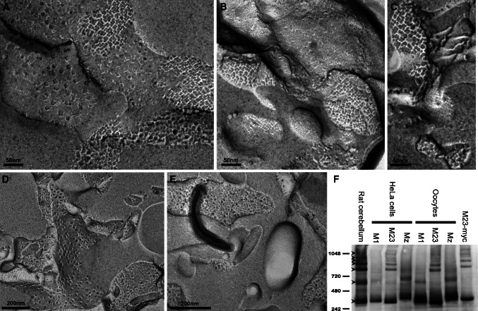
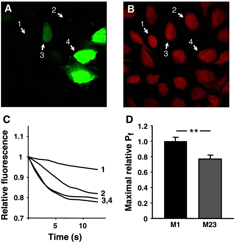
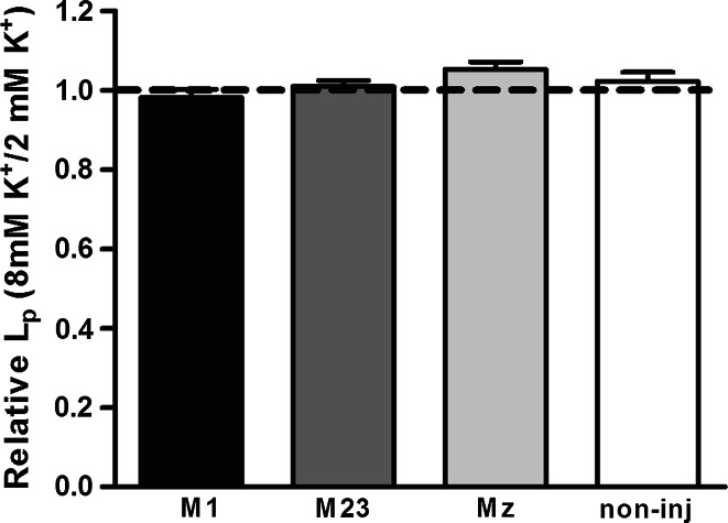
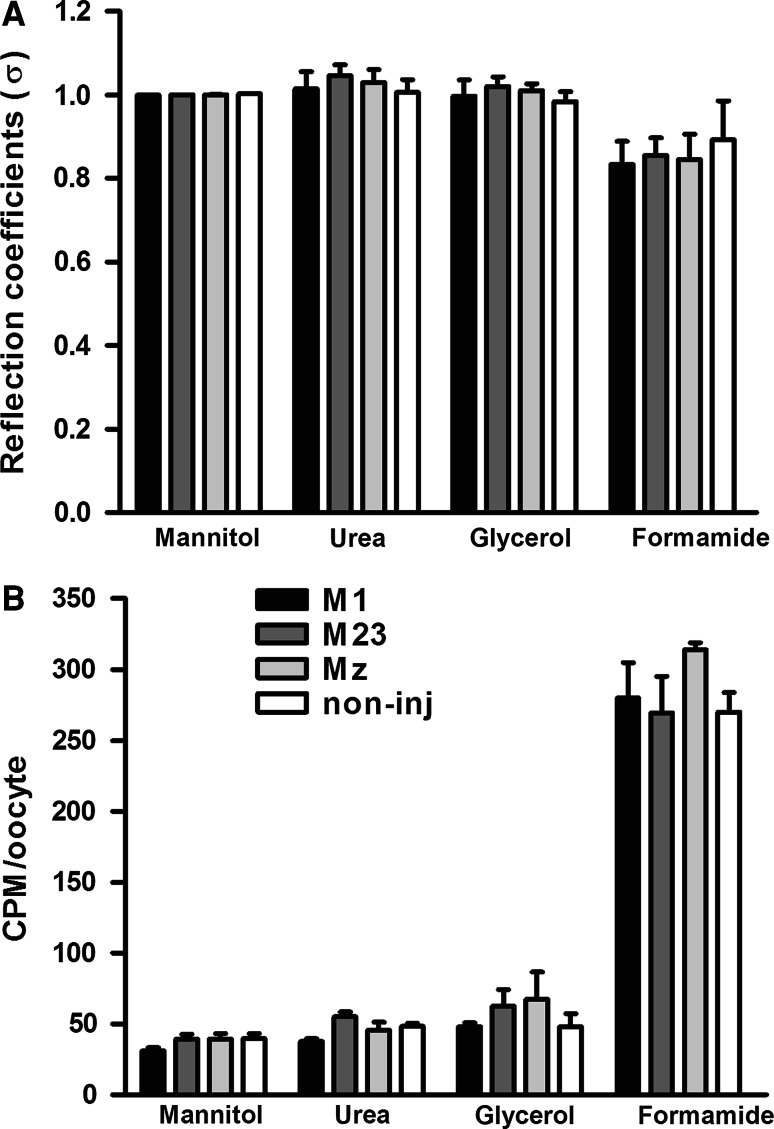
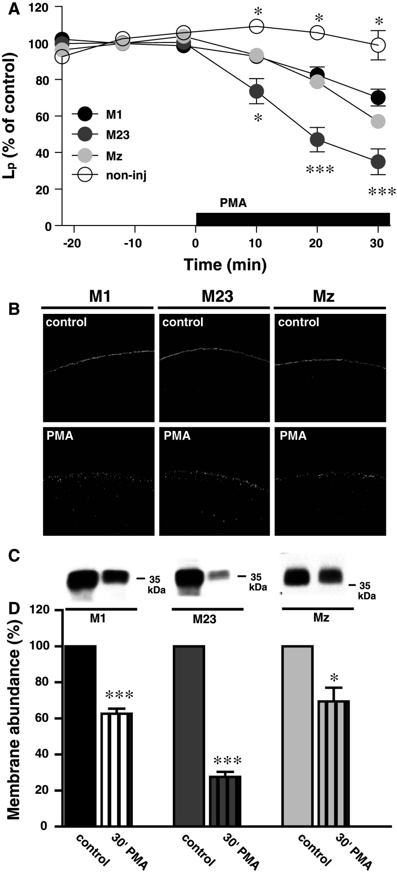
Aquaporin 4 (AQP4) is expressed in the perivascular glial endfeet and is an important pathway for water during formation and resolution of brain edema. In this study, we examined the functional properties and relative unit water permeability of three functional isoforms of AQP4 expressed in the brain (M1, M23, Mz). The M23 isoform gave rise to square arrays when expressed in Xenopus laevis oocytes. The relative unit water permeability differed significantly between the isoforms in the order of M1 > Mz > M23. None of the three isoforms were permeable to small osmolytes nor were they affected by changes in external K(+) concentration. Upon protein kinase C (PKC) activation, oocytes expressing the three isoforms demonstrated rapid reduction of water permeability, which correlated with AQP4 internalization. The M23 isoform was more sensitive to PKC regulation than the longer isoforms and was internalized significantly faster. Our results suggest a specific role for square array formation.


Figures

Fig. 1
Fig. 1
The M23 isoform forms square arrays in Xenopus laevis oocytes. ac At the plasma membrane, highly ordered structures characteristic of square arrays are observed for the M23 isoform. In contrast, neither the M1 isoform (d) nor the Mz isoform (e) show similar ordered structures. f BN-PAGE analysis of AQP4. In contrast to the M1 and Mz isoforms, the M23 isoform forms higher molecular weight moieties in both HeLa cells and Xenopus oocytes. Lane 1 brain lysate (rat cerebellum), lanes 2–4 M1, M23, and Mz expressed in HeLa cells, lanes 5–7 M1, M23, and Mz expressed in Xenopus oocytes. Control M23-myc expressed in HeLa cells also exhibited higher order bands (lane 8). Molecular weight markers are indicated to the left, in kDa. The lower tetramer band and six higher order bands are indicated by arrowheads
Fig. 2
Fig. 2
The relative unit water permeability of M1, M23, and Mz expressed in oocytes. a An oocyte expressing the M23 isoform and a non-injected oocyte (with L ps of 1.14 and 0.11 × 10−3 cm/s, respectively) were challenged with an osmotic gradient of 20 mOsm mannitol for 30 s. b The average water permeability of oocytes expressing M1, M23, or Mz with the contribution of the native oocyte membrane deducted (n = 20–25 of each). c Representative confocal laser scanning microscopy of oocytes expressing M1, M23 and Mz immunolabeled for AQP4. d Oocyte total fluorescent counts (in arbitrary units) were used to asses the abundance of AQP4 in oocytes expressing the three isoforms (n = 15–20 of each). e Oocyte plasma membrane fluorescent counts (in arbitrary units) were used to asses the AQP4 abundance in the plasma membrane of oocytes expressing the three isoforms (n = 15–20 of each). f Normalized relative unit water permeability of the three isoforms based on quantification by immunocytochemistry (n = 4–5 experiments based on 3–5 oocytes of each). g Normalized relative unit water permeability of the three isoforms based on quantification by immunoblotting of purified plasma membranes (n = 3 experiments, each based on n = 5 oocytes of each isoform for the L p determination and n = 20 oocytes of each isoform for the plasma membrane purification). ***P < 0.001 (compared to M1)
Fig. 3
Fig. 3
The relative unit water permeability of M1 and M23 in transfected human bronchial epithelial cell line. a The level of GFP fluorescence within the cells. b Loading with calcein, which was used for water permeability measurements, was similar in AQP4-positive and AQP4-negative cells. Numbers indicate the same cells before (a) and after (b) calcein loading. c Single cell traces showing the dilution of calcein due to the cell swelling after an osmotic challenge. In cells with low GFP (and hence AQP4) expression (cells 1–3 in a), the swelling rate was increasing (line 1 through line 3 in c) with the increase in GFP (AQP4) level. The swelling of the cells 3 and 4 occurred at practically identical speed, probably due to saturation of the plasma membrane with AQP4. d Maximal relative water permeability in cells expressing M1 (n = 22) and M23 (n = 13), **P < 0.01
Fig. 4
Fig. 4
Lack of K+-dependent water permeability in M1, M23, and Mz-expressing oocytes. Oocytes expressing the three different isoforms were voltage-clamped to −50 mV to avoid K+-dependent changes in membrane potential and exposed to test solution containing the standard 2 mM K+ (+6 mM Ch+) in which the L p was determined. Subsequently the same oocyte was exposed to a test solution containing 8 mM K+ in which the L p was determined. The data are presented as the L p obtained in 8 mM K+ relative to that obtained in 2 mM K+ (n = 4 of each)
Fig. 5
Fig. 5
Lack of permeability to small osmolytes in M1, M23, and Mz-expressing oocytes. a The L p of oocytes expressing M1, M23, and Mz as well as non-injected oocytes was determined with different osmolytes; mannitol, urea, glycerol, and formamide (20 mOsm of each). The L p obtained with urea, glycerol, and formamide was plotted relative to that obtained with mannitol for each oocyte (reflection coefficient, σ). The reflection coefficients for oocytes expressing M1, M23, or Mz were not significantly different from that of the non-injected oocytes (n = 4 of each isoform and n = 3 for the non-injected oocytes). b Oocytes expressing M1, M23, and Mz as well as non-injected oocytes were exposed to test solution containing different osmolytes; mannitol, urea, glycerol, and formamide (20 mOsm of each) in addition to trace amounts of the [14C]-labeled osmolyte. The data are presented as the average uptake of four experiments performed in pentaplicate, with no significant difference between oocytes expressing the three isoforms and the non-injected oocytes
Fig. 6
Fig. 6
PKC-dependent down-regulation of AQP4. a The relative water permeability of oocytes expressing M1, M23, or Mz or non-injected oocytes as a function of time. 1 nM PMA was included in the external solution as marked by the black bar. After 30 min of PMA treatment, the L p was reduced to (in % of control) for M1; 70 ± 5 (n = 8), M23; 35 ± 7 (n = 7), Mz; 57 ± 4, error bar within the symbol (n = 9), and non-injected; 99 ± 8 (n = 3). The significance levels on the graph refer to M1, *P < 0.05, ***P < 0.001. b Representative confocal laser scanning microscopy of oocytes expressing M1, M23, and Mz immunolabeled for AQP4 without (upper panels) or with (lower panels) 30 min PMA treatment (1 nM). c Representative immunoblot of plasma membrane purification of oocytes expressing M1, M23, or Mz in control condition and after 30 min of PMA treatment (1 nM), minimum 15 oocytes for each condition. d Relative membrane abundance of M1, M23, and Mz was assessed by densitometry of the immunoblots as presented in panel (c) of oocytes with control treatment or 30 min PMA treatment (1 nM). 15–20 oocytes were used for each condition (n = 3 experiments), *P < 0.05, ***P < 0.001. The M23-expressing oocytes had a significantly lower AQP4 abundance in the plasma membrane after PMA treatment compared to M1 and Mz, P < 0.01

References

    1. Amiry-Moghaddam M, Ottersen OP. The molecular basis of water transport in the brain. Nat Rev Neurosci. 2003;4:991–1001. doi: 10.1038/nrn1252. - DOI - PubMed
    1. Nielsen S, Nagelhus EA, Amiry-Moghaddam M, Bourque C, Agre P, Ottersen OP. Specialized membrane domains for water transport in glial cells: high-resolution immunogold cytochemistry of aquaporin-4 in rat brain. J Neurosci. 1997;17:171–180. - PMC - PubMed
    1. Noell S, Fallier-Becker P, Beyer C, Kroger S, Mack AF, Wolburg H. Effects of agrin on the expression and distribution of the water channel protein aquaporin-4 and volume regulation in cultured astrocytes. Eur J Neurosci. 2007;26:2109–2118. doi: 10.1111/j.1460-9568.2007.05850.x. - DOI - PubMed
    1. Zador Z, Stiver S, Wang V, Manley GT. Role of aquaporin-4 in cerebral edema and stroke. Handb Exp Pharmacol. 2009;190:159–170. doi: 10.1007/978-3-540-79885-9_7. - DOI - PMC - PubMed
    1. Carmosino M, Procino G, Tamma G, Mannucci R, Svelto M, Valenti G. Trafficking and phosphorylation dynamics of AQP4 in histamine-treated human gastric cells. Biol Cell. 2007;99:25–36. doi: 10.1042/BC20060068. - DOI - PubMed

Publication types

LinkOut - more resources