. 2010 Jul;169(7):801-11.
doi: 10.1007/s00431-009-1115-9. Epub 2009 Dec 15.

Schimke immunoosseous dysplasia: defining skeletal features

Affiliations

Schimke immunoosseous dysplasia: defining skeletal features

Kshamta B Hunter et al. Eur J Pediatr. 2010 Jul.

Abstract

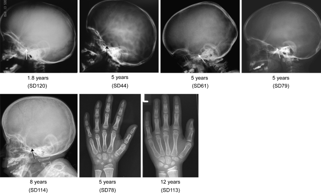
Schimke immunoosseous dysplasia (SIOD) is an autosomal recessive multisystem disorder characterized by prominent spondyloepiphyseal dysplasia, T cell deficiency, and focal segmental glomerulosclerosis. Biallelic mutations in swi/snf-related, matrix-associated, actin-dependent regulator of chromatin, subfamily a-like 1 (SMARCAL1) are the only identified cause of SIOD, but approximately half of patients referred for molecular studies do not have detectable mutations in SMARCAL1. We hypothesized that skeletal features distinguish between those with or without SMARCAL1 mutations. Therefore, we analyzed the skeletal radiographs of 22 patients with and 11 without detectable SMARCAL1 mutations. We found that patients with SMARCAL1 mutations have a spondyloepiphyseal dysplasia (SED) essentially limited to the spine, pelvis, capital femoral epiphyses, and possibly the sella turcica, whereas the hands and other long bones are basically normal. Additionally, we found that several of the adolescent and young adult patients developed osteoporosis and coxarthrosis. Of the 11 patients without detectable SMARCAL1 mutations, seven had a SED indistinguishable from patients with SMARCAL1 mutations. We conclude therefore that SED is a feature of patients with SMARCAL1 mutations and that skeletal features do not distinguish who of those with SED have SMARCAL1 mutations.


Figures

Fig. 1
Fig. 1
Typical bony features of SIOD. a Lateral spine radiograph of a 5-year-old child showing dorsally flattened, pear-shaped vertebral bodies. b Lateral skull radiograph of a 5-year-old child showing the typical widening of the sella. c Posteroanterior hand radiograph of a 13-year-old adolescent showing the absence of bony abnormalities. d Anteroposterior hip radiograph of a 4-year-old child showing the small, laterally displaced capital femoral epiphyses, hypoplastic basilar ilia, and upslanting and poorly formed acetabula
Fig. 2
Fig. 2
Lateral spine radiographs of patients with identified SMARCAL1 mutations at different ages. The vertebral bodies are flattened at all ages and pear-shaped between 4 and 6 years. There is generalized vertebral flattening with slightly irregular upper and lower plates in some patients. The severity of the vertebral changes is variable as illustrated by comparison of SD112 with SD66. Note the radiolucency of adult vertebrae consistent with the progressive osteopenia of SIOD patients. SD27 had diffuse disc calcification; the significance of this is uncertain
Fig. 3
Fig. 3
Anterior–posterior spine radiographs of patients with identified SMARCAL1 mutations at different ages show various degrees of platyspondyly. SD27 had diffuse disc calcification; the significance of this is uncertain
Fig. 4
Fig. 4
Hip radiographs of patients with identified SMARCAL1 mutations at different ages. The femora generally have small, laterally displaced capital epiphyses, but normal epiphyseal ossification may occur (e.g., SD78, SD 112). By adulthood, the femoral dysplasia usually progresses to premature coxarthrosis (SD18 and SD27) requiring prosthetic therapy (SD27). The ilia are usually small because of hypoplastic basilar portions and have upslanting and poorly formed acetabula. The severity of the ilia and femoral changes is variable as illustrated by comparison of SD78, who had preserved capital femoral epiphyses and a normal pelvis, with SD121, who had short femoral necks with a peculiar lip-like medial protrusion and markedly hypoplastic basilar ilia
Fig. 5
Fig. 5
Skull and hand radiographs of patients with identified SMARCAL1 mutations at different ages. Note the marked widening of the sella in SD44, SD61, and SD79 and mild widening of the sella in SD120. No bony abnormalities are observed in the hands
Fig. 6
Fig. 6
Lateral spine and hip radiographs of SIOD patients without detectable SMARCAL1 mutations at different ages. Note the similarity of bony features to those with SMARCAL1 mutations. SD54 had kyphoscoliosis and anterior hypoplasia of L2, which may have been secondary to an unrelated muscular hypotonia

References

    1. Bansbach CE, Betous R, Lovejoy CA, et al. The annealing helicase SMARCAL1 maintains genome integrity at stalled replication forks. Genes Dev. 2009;23:2405–2414. doi: 10.1101/gad.1839909. - DOI - PMC - PubMed
    1. Baradaran-Heravi A, Thiel C, Rauch A, et al. Clinical and genetic distinction of Schimke immuno-osseous dysplasia and cartilage-hair hypoplasia. Am J Med Genet A. 2008;146A:2013–2017. doi: 10.1002/ajmg.a.32406. - DOI - PMC - PubMed
    1. Boerkoel CF, O’Neill S, Andre JL, et al. Manifestations and treatment of Schimke immuno-osseous dysplasia: 14 new cases and a review of the literature. Eur J Pediatr. 2000;159:1–7. doi: 10.1007/s004310050001. - DOI - PubMed
    1. Boerkoel CF, Takashima H, John J, et al. Mutant chromatin remodeling protein SMARCAL1 causes Schimke immuno-osseous dysplasia. Nat Genet. 2002;30:215–220. doi: 10.1038/ng821. - DOI - PubMed
    1. Cheung VG, Spielman RS. Genetics of human gene expression: mapping DNA variants that influence gene expression. Nat Rev Genet. 2009;10:595–604. doi: 10.1038/nrg2630. - DOI - PMC - PubMed

Publication types