

Oxidative lipidomics of apoptosis: quantitative assessment of phospholipid hydroperoxides in cells and tissues
- PMID: 20013189
- PMCID: PMC6746671
- DOI: 10.1007/978-1-60327-029-8_21
Oxidative lipidomics of apoptosis: quantitative assessment of phospholipid hydroperoxides in cells and tissues
Abstract
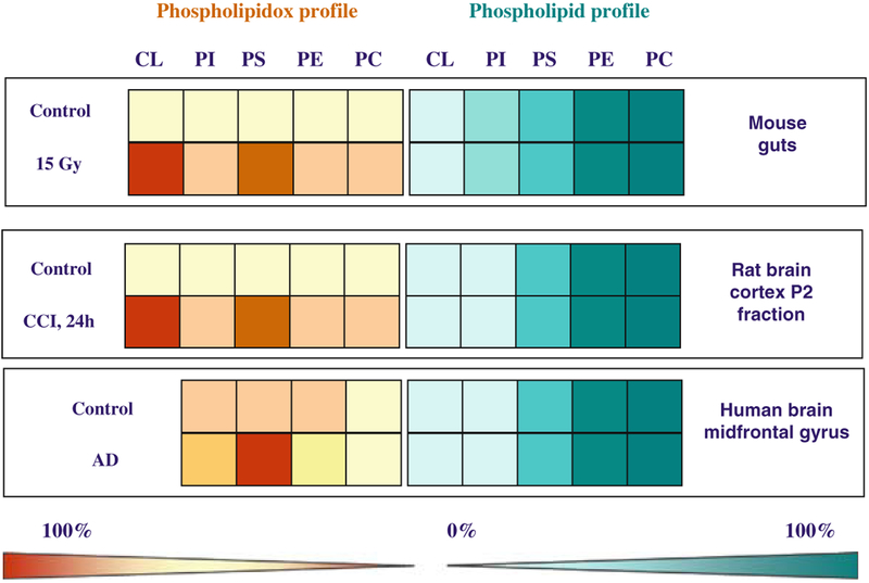
Oxidized phospholipids play essential roles in execution of mitochondrial stage of apoptosis and clearance of apoptotic cells. The identification and quantification of oxidized phospholipids generated during apoptosis can be successfully achieved by oxidative lipidomics. With this approach, diverse molecular species of phospholipids and their hydroperoxides are identified and characterized by soft-ionization mass-spectrometry techniques such as electrospray ionization (ESI). Quantitative assessment of lipid hydroperoxides is performed by fluorescence HPLC-based protocol. The protocol is based on separation of phospholipids using two-dimensional-high-performance thin-layer chromatography (2-D-HPTLC). Phospholipids are hydrolyzed using phospholipase A(2). The fatty acid hydroperoxides (FA-OOH) released is quantified by a fluorometric assay using Amplex red reagent and microperoxidase-11 (MP-11). Detection limit of this protocol is 1-2 pmol of lipid hydroperoxides. Lipid arrays vs. oxidized lipid arrays can be performed by comparing the abundance of phospholipids with the abundance of oxidized phospholipids. Using oxidative lipidomics approach we show that the pattern of phospholipid oxidation during apoptosis is nonrandom and does not follow their abundance in several types of cells undergoing apoptosis and a variety of disease states. This has important implications for evaluation of apoptosis in vivo. The anionic phospholipids, cardiolipin (CL) and phosphatidylserine (PS), are the preferred peroxidation substrates.
Figures
Similar articles
-
Oxidative lipidomics of programmed cell death.Methods Enzymol. 2008;442:375-93. doi: 10.1016/S0076-6879(08)01419-5. Methods Enzymol. 2008. PMID: 18662580
-
Mass-spectrometric characterization of phospholipids and their hydroperoxide derivatives in vivo: effects of total body irradiation.Methods Mol Biol. 2009;580:153-83. doi: 10.1007/978-1-60761-325-1_9. Methods Mol Biol. 2009. PMID: 19784599
-
Mass-spectrometric analysis of hydroperoxy- and hydroxy-derivatives of cardiolipin and phosphatidylserine in cells and tissues induced by pro-apoptotic and pro-inflammatory stimuli.J Chromatogr B Analyt Technol Biomed Life Sci. 2009 Sep 15;877(26):2863-72. doi: 10.1016/j.jchromb.2009.03.007. Epub 2009 Mar 13. J Chromatogr B Analyt Technol Biomed Life Sci. 2009. PMID: 19328050 Free PMC article.
-
Oxidized phosphatidylserine: production and bioactivities.Yonago Acta Med. 2014 Dec;57(4):119-27. Epub 2014 Dec 26. Yonago Acta Med. 2014. PMID: 25901098 Free PMC article. Review.
-
Lipid hydroperoxides as a source of singlet molecular oxygen.Subcell Biochem. 2014;77:3-20. doi: 10.1007/978-94-007-7920-4_1. Subcell Biochem. 2014. PMID: 24374914 Review.
Cited by
-
Intraoral Mitochondrial-Targeted GS-Nitroxide, JP4-039, Radioprotects Normal Tissue in Tumor-Bearing Radiosensitive Fancd2(-/-) (C57BL/6) Mice.Radiat Res. 2016 Feb;185(2):134-50. doi: 10.1667/RR14035.1. Epub 2016 Jan 20. Radiat Res. 2016. PMID: 26789701 Free PMC article.
-
Cardiolipin asymmetry, oxidation and signaling.Chem Phys Lipids. 2014 Apr;179:64-9. doi: 10.1016/j.chemphyslip.2013.11.010. Epub 2013 Dec 1. Chem Phys Lipids. 2014. PMID: 24300280 Free PMC article. Review.
-
Therapies targeting lipid peroxidation in traumatic brain injury.Brain Res. 2016 Jun 1;1640(Pt A):57-76. doi: 10.1016/j.brainres.2016.02.006. Epub 2016 Feb 10. Brain Res. 2016. PMID: 26872597 Free PMC article. Review.
-
Mass-spectrometric characterization of peroxidized and hydrolyzed lipids in plasma and dendritic cells of tumor-bearing animals.Biochem Biophys Res Commun. 2011 Sep 16;413(1):149-53. doi: 10.1016/j.bbrc.2011.08.074. Epub 2011 Aug 22. Biochem Biophys Res Commun. 2011. PMID: 21872574 Free PMC article.
-
Relationship Between Anti-DFS70 Autoantibodies and Oxidative Stress.Biomark Insights. 2022 Jan 30;17:11772719211066791. doi: 10.1177/11772719211066791. eCollection 2022. Biomark Insights. 2022. PMID: 35125863 Free PMC article.
References
-
- Vijg J and Suh Y (2005) Genetics of longevity and aging. Annu. Rev. Med 56, 193–212. - PubMed
-
- Watson AD (2006) Thematic review series: systems biology approaches to metabolic and cardiovascular disorders. Lipidomics: a global approach to lipid analysis in biological systems. J. Lipid Res 47, 2101–2111. - PubMed
-
- Gaspar ML, Aregullin MA, Jesch SA, Nunez LR, Villa-García M, and Henry SA (2007) The emergence of yeast lipidomics. Biochim. Biophys. Acta 1771, 241–254. - PubMed
-
- Marathe GK, Harrison KA, Murphy RC, Prescott SM, Zimmerman GA, and McIntyre TM (2000) Bioactive phospholipid oxidation products. Free Radic. Biol. Med 28, 1762–1770. - PubMed
-
- DeLong CJ, Baker PR, Samuel M, Cui Z, and Thomas MJ (2001) Molecular species composition of rat liver phospholipids by ESI-MS/MS: the effect of chromatography. J. Lipid Res 42, 1959–1968. - PubMed
Publication types
MeSH terms
Substances
Grants and funding
LinkOut - more resources
Full Text Sources
