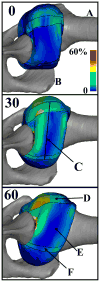
Finite element modelling of the glenohumeral capsule can help assess the tested region during a clinical exam
- PMID: 20013435
- PMCID: PMC3769951
- DOI: 10.1080/10255840903317378
Finite element modelling of the glenohumeral capsule can help assess the tested region during a clinical exam
Abstract
The objective of this research was to examine the efficacy of evaluating the region of the glenohumeral capsule being tested by clinical exams for shoulder instability using finite element (FE) models of the glenohumeral joint. Specifically, the regions of high capsule strain produced by glenohumeral joint positions commonly used during a clinical exam were identified. Kinematics that simulated a simple translation test with an anterior load at three external rotation angles were applied to a validated, subject-specific FE model of the glenohumeral joint at 60° of abduction. Maximum principal strains on the glenoid side of the inferior glenohumeral ligament (IGHL) were significantly higher than the maximum principal strains on the humeral side, for all three regions of the IGHL at 30° and 60° of external rotation. These regions of localised strain indicate that these joint positions might be used to test the glenoid side of the IGHL during this clinical exam, but are not useful for assessing the humeral side of the IGHL. The use of FE models will facilitate the search for additional joint positions that isolate high strains to other IGHL regions, including the humeral side of the IGHL.
Figures


References
-
- Bankart ASB. The pathology and treatment of recurrent dislocation of the shoulder joint. Br J Surg. 1938;26:23–9.
-
- Bigliani LU, Pollock RG, Soslowsky LJ, Flatow EL, Pawluk RJ, Mow VC. Tensile properties of the inferior glenohumeral ligament. J Orthop Res. 1992;10:187–97. - PubMed
-
- Bokor DJ, Conboy VB, Olson C. Anterior instability of the glenohumeral joint with humeral avulsion of the glenohumeral ligament. A review of 41 cases. J Bone Joint Surg Br. 1999;81:93–6. - PubMed
-
- Brenneke SL, Reid J, Ching RP, Wheeler DL. Glenohumeral kinematics and capsulo-ligamentous strain resulting from laxity exams. Clin Biomech (Bristol, Avon) 2000;15:735–42. - PubMed
Publication types
MeSH terms
Grants and funding
LinkOut - more resources
Full Text Sources
