. 2010;54(1):175-80.
doi: 10.1387/ijdb.082610bl.

Signalling molecules involved in mouse bladder smooth muscle cellular differentiation

Affiliations

Signalling molecules involved in mouse bladder smooth muscle cellular differentiation

Benchun Liu et al. Int J Dev Biol. 2010.

Abstract

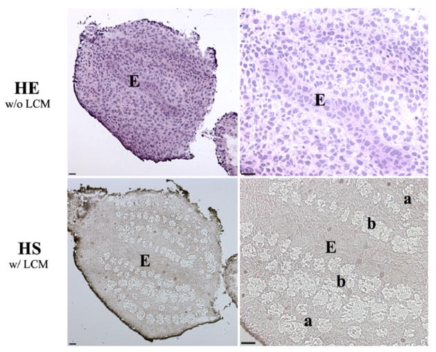
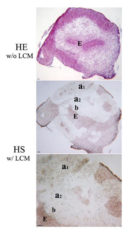
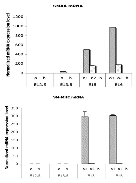
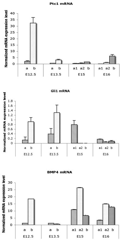
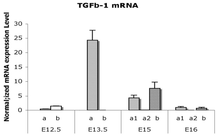
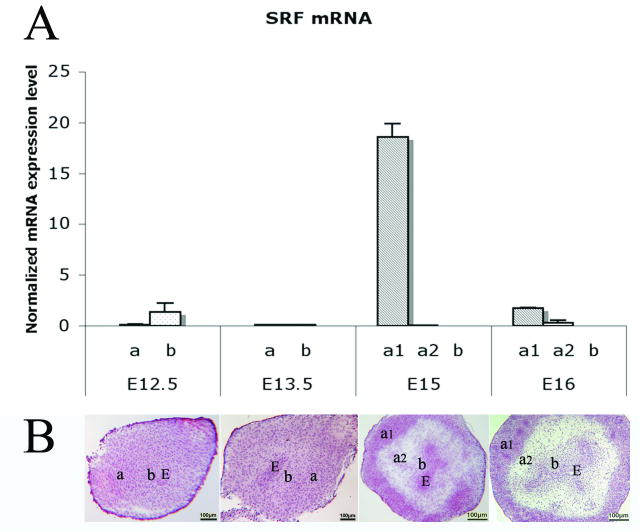
Mouse bladder mesenchyme differentiates into smooth muscle under the influence of urothelium at gestational day 13.5 (E13.5). Sonic hedgehog (Shh) is considered to be the upstream gene arising from the urothelium, which induces smooth muscle in the peripheral bladder mesenchyme. We hypothesize differential gene expression across the full thickness of bladder mesenchyme as a function of proximity to the inducing bladder urothelium and the peripheral location of the smooth muscle. Embryonic bladders from FVB mice were collected at E12.5, 13.5, 15 and 16 and cryosectioned followed by microdissection with a PixCell II laser capture microscope. RNA extraction was performed at the laser captured sites and mRNA expression profiles were measured using SYBR Green quantitative RT-PCR. Smooth muscle a-actin (SMAA) and smooth muscle myosin heavy chain (SM-MHC) were expressed in the E13.5, E15 and E16 bladders in the peripheral layer of mesenchyme, but not in the prospective submucosa. Patched 1 (Ptc1), Gli1 and bone morphogenetic protein (Bmp) 4 expression was consistently elevated in the mesenchymal layer immediately adjacent to the urothelium compared to the peripheral location at E12.5. After E12.5, Ptc1 expression decreased to an undetectable level throughout the bladder mesenchyme. The level of TGF-beta1 was highest in the mesenchymal layer adjacent to the serosa at E13.5. The level of expression of serum response factor (SRF) was also highest at E15 in the peripheral mesenchyme. Genes downstream of Shh are differentially expressed in the prospective submucosa vs. the peripheral bladder mesenchyme as a function gestation age and smooth muscle differentiation.


Figures

Fig. 1
Fig. 1. Laser-assisted microdissection of mesenchyme in the E13.5 mouse bladder. (HE w/o LCM)
Morphology of HE staining on serial section for laser capture microdissection. (HS w/ LCM) Morphology of HS staining after laser capture microdissection. Mesenchymal locations were chosen either immediately adjacent to the urothelium in the prospective submucosa or next to the serosal layer in the peripheral bladder mesenchyme, an area of future smooth muscle differentiation in E12.5 and E13.5 bladders. a, Serosal zone. b, Submucosal zone. E, Epithelium. Scale bar, 50μm.
Fig. 2
Fig. 2. Laser-assisted microdissection of mesenchyme in the E15 mouse bladder. (HE w/o LCM)
Morphology of HE staining on serial section for laser capture microdissection. (HS w/ LCM) Morphology of HS staining after laser capture microdissection. Bladder mesenchymes were captured in the submucosa and in the area of peripheral smooth muscle in E15 and E16. a1, Smooth muscle layer. a2, Intermediate zone. b, Submucosal zone. E, Epithelium. Scale bar, 50μm.
Fig. 3
Fig. 3. Real time RT-PCR analysis on mRNA expressions of SMAA and SM-MHC in laser captured mesenchymal components of each gestational stage
Expression of SMAA mRNA was detectable in the peripheral cellular population at E13.5 and increased to significantly high levels in smooth muscle cells at E15 and E16. SM-MHC was detectable temporal slightly later than SMAA and increasing in expression at E15 and E16 (Mann-Whitney test, p<0.001). a, Serosal zone. a1, Smooth muscle layer. a2, Intermediate zone. b, Submucosal zone.
Fig. 4
Fig. 4. Real time RT-PCR analysis on mRNA expressions of Ptc1, Gli1, and Bmp4 in laser captured mesenchymal components of each gestational stage
Ptc1, Gli1, and Bmp4. expression were restricted to a thin layer of mesenchymal cells in the submucosa immediately adjacent to the epithelium. Both Ptc1 and Bmp4 genes were significantly decreased in all mesenchymal cells in the E13.5 bladder. Ptc1 remained at a low level in contrast to Bmp4 in which was increased at E15 and E16. Gli1 gene expression was up regulated in the submucosa at E13.5. At E15 the expression changed to the smooth muscle layer (Mann-Whitney test, p<0.001). a, Serosal zone. a1, Smooth muscle layer. a2, Intermediate zone. b, Submucosal zone.
Fig. 5
Fig. 5. Real time RT-PCR analysis on mRNA expression of TGF-β1 in laser captured mesenchymal components of each gestational stage
TGF-β1 expression was minimal in the E12.5 bladder mesenchyme. TGF-β1 increased to the highest level in the smooth muscle differentiation layer adjacent to the serosa at E13.5 and then decreased in all mesenchymal compartments in the E15 and E16 (Mann-Whitney test, p<0.001). a, Serosal zone. a1, Smooth muscle layer. a2, Intermediate zone. b, Submucosal zone.
Fig. 6
Fig. 6. Real time RT-PCR analysis on mRNA expression of SRF in laser captured mesenchymal components of each gestational stage and mouse bladder developmental illustrations
(A) SRF was significantly up regulated in smooth muscle cells at E15 compared to other mesenchymal components at E12.5, E13.5, and E16 (Mann-Whitney test, p<0.001). (B) Mouse bladder developmental illustrations with HE staining show locations of a1, a2 and b nomenclatures. a, Serosal zone. a1, Smooth muscle layer. a2, Intermediate zone. b, Submucosal zone. E, Bladder epithelium. Scale bar, 100μm.

Similar articles

Cited by

References

    1. Airik R, Bussen M, Singh MK, Petry M, Kispert A. Tbx18 regulates the development of the ureteral mesenchyme. J Clin Invest. 2006;116:663–74. - PMC - PubMed
    1. Baskin LS, Hayward SW, Young P, Cunha GR. Role of mesenchymal–epithelial interactions in normal bladder development. J Urol. 1996;156:1820–7. - PubMed
    1. Bonner RF, Emmert-Buck M, Cole K, Pohida T, Chuagui R, Goldstein S, Littoa LA. Laser capture microdissection: molecular analysis of tissue. Science. 1997;278:1481–1483. - PubMed
    1. Bragg AD, Moses HL, Serra R. Signaling to the epithelium is not sufficient to mediate all of the effects of transforming growth factor beta and bone morphogenetic protein 4 on murine embryonic lung development. Mech Dev. 2001;109:13–26. - PubMed
    1. Browning CL, Culberson DE, Aragon IV, Fillmore RA, Croissant JD, Schwartz RJ, ZiImmer WE. The developmentally regulated expression of serum response factor plays a key role in the control of smooth muscle-specific genes. Dev Biol. 1998;194:18–37. - PubMed

Publication types

MeSH terms