

A novel role of gap junction connexin46 protein to protect breast tumors from hypoxia
- PMID: 20013805
- PMCID: PMC3150590
- DOI: 10.1002/ijc.25107
A novel role of gap junction connexin46 protein to protect breast tumors from hypoxia
Abstract
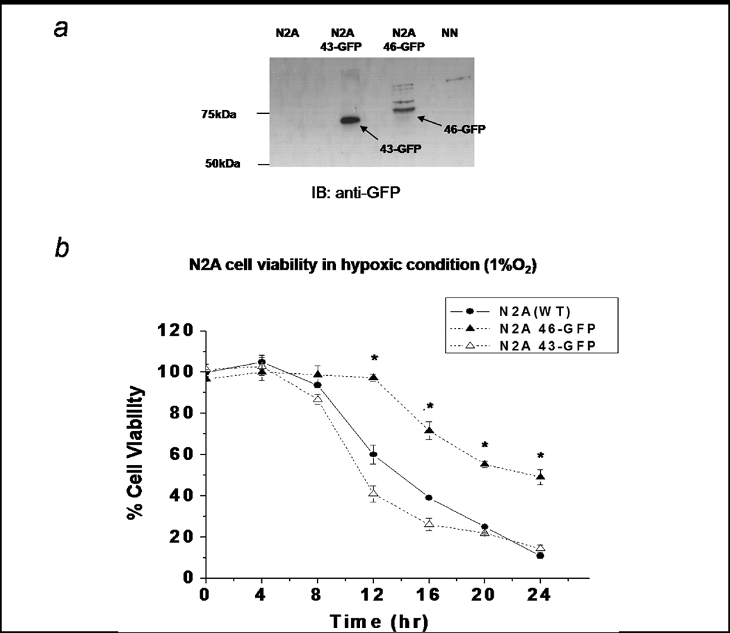
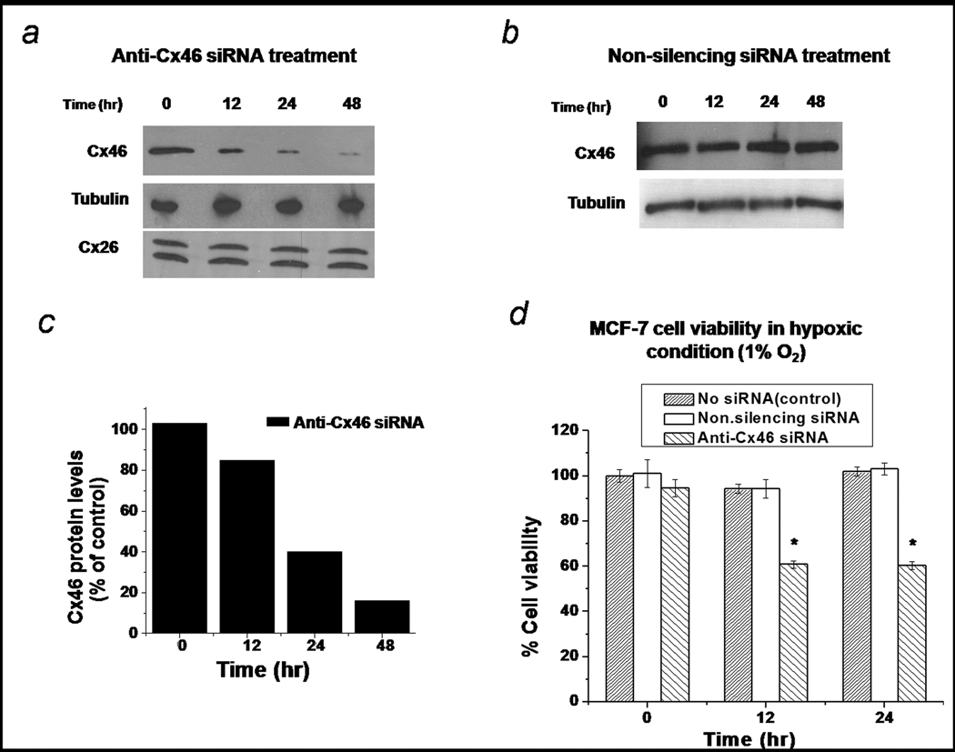
Connexin proteins are the principle structural components of the gap junctions. Colocalization and tissue-specific expression of diverse connexin molecules are reported to occur in a variety of organs. Impairment of gap junctional intercellular communication, caused by mutations, gain of function or loss of function of connexins, is involved in a number of diseases including the development of cancer. Here we show that human breast cancer cells, MCF-7 and breast tumor tissues express a novel gap junction protein, connexin46 (Cx46) and it plays a critical role in hypoxia. Previous studies have shown that connexin46 is predominantly expressed in lens and our studies find that Cx46 protects human lens epithelial cells from hypoxia induced death. Interestingly, we find that Cx46 is upregulated in MCF-7 breast cancer cells and human breast cancer tumors. Downregulation of Cx46 by siRNA promotes 40% MCF-7 cell death at 24 hr under hypoxic conditions. Furthermore, direct injection of anti-Cx46 siRNA into xenograft tumors prevents tumor growth in nude mice. This finding will provide an exciting new direction for drug development for breast cancer treatment and suggests that both normal hypoxic tissue (lens) and adaptive hypoxic tissue (breast tumor) utilize the same protein, Cx46, as a protective strategy from hypoxia.
Figures


Similar articles
-
Investigation of the reciprocal relationship between the expression of two gap junction connexin proteins, connexin46 and connexin43.J Biol Chem. 2011 Jul 8;286(27):24519-33. doi: 10.1074/jbc.M110.217208. Epub 2011 May 23. J Biol Chem. 2011. PMID: 21606502 Free PMC article.
-
Connexins in Cancer, the Possible Role of Connexin46 as a Cancer Stem Cell-Determining Protein.Biomolecules. 2023 Sep 27;13(10):1460. doi: 10.3390/biom13101460. Biomolecules. 2023. PMID: 37892142 Free PMC article. Review.
-
Treatment with connexin 46 siRNA suppresses the growth of human Y79 retinoblastoma cell xenografts in vivo.Exp Eye Res. 2011 Apr;92(4):251-9. doi: 10.1016/j.exer.2011.02.003. Epub 2011 Feb 12. Exp Eye Res. 2011. PMID: 21320488 Free PMC article.
-
Connexin46 is retained as monomers in a trans-Golgi compartment of osteoblastic cells.J Cell Biol. 1997 May 19;137(4):847-57. doi: 10.1083/jcb.137.4.847. J Cell Biol. 1997. PMID: 9151687 Free PMC article.
-
Regulation of Connexin Gap Junctions and Hemichannels by Calcium and Calcium Binding Protein Calmodulin.Int J Mol Sci. 2020 Nov 2;21(21):8194. doi: 10.3390/ijms21218194. Int J Mol Sci. 2020. PMID: 33147690 Free PMC article. Review.
Cited by
-
The potential prognostic value of connexin 26 and 46 expression in neoadjuvant-treated breast cancer.BMC Cancer. 2013 Feb 2;13:50. doi: 10.1186/1471-2407-13-50. BMC Cancer. 2013. PMID: 23374644 Free PMC article.
-
Prognostic value of the mRNA expression of gap junction α members in patients with gastric cancer.Oncol Lett. 2019 Aug;18(2):1669-1678. doi: 10.3892/ol.2019.10516. Epub 2019 Jun 21. Oncol Lett. 2019. PMID: 31423234 Free PMC article.
-
Distinctive actions of connexin 46 and connexin 50 in anterior pituitary folliculostellate cells.PLoS One. 2017 Jul 31;12(7):e0182495. doi: 10.1371/journal.pone.0182495. eCollection 2017. PLoS One. 2017. PMID: 28759642 Free PMC article.
-
Complementary expression and phosphorylation of Cx46 and Cx50 during development and following gene deletion in mouse and in normal and orchitic mink testes.Am J Physiol Regul Integr Comp Physiol. 2015 Aug 1;309(3):R255-76. doi: 10.1152/ajpregu.00152.2015. Epub 2015 May 27. Am J Physiol Regul Integr Comp Physiol. 2015. PMID: 26017495 Free PMC article.
-
Investigation of the reciprocal relationship between the expression of two gap junction connexin proteins, connexin46 and connexin43.J Biol Chem. 2011 Jul 8;286(27):24519-33. doi: 10.1074/jbc.M110.217208. Epub 2011 May 23. J Biol Chem. 2011. PMID: 21606502 Free PMC article.
References
-
- Goodenough D, Goliger J, Paul D. Connexins, connexons, and intercellular communication. Annu Rev Biochem. 1996;65:475–502. - PubMed
-
- Loewenstein W. Junctional intercellular communication and the control of growth. Biochim Biophys Acta. 1979;560:1–65. - PubMed
-
- Trosko JE, Ruch RJ. Cell-Cell Communication in carcinogenesis. Front. Biosci. 1998;3:208–236. - PubMed
-
- Kanczuga-Koda L, Sulkowska M, Koda M, Reszec J, Famulski W, Baltaziak M, Sulkowski S. Expression of connexin 43 in breast cancer in comparison with mammary dysplasia and the normal mammary gland. Folia Morphol (Warsz) 2003;62:439–442. - PubMed
Publication types
MeSH terms
Substances
Grants and funding
LinkOut - more resources
Full Text Sources
Medical
Miscellaneous
