

doi: 10.1002/anie.200905483.
Multiple base-recognition sites in a biological nanopore: two heads are better than one
Affiliations
- PMID: 20014084
- PMCID: PMC3128935
- DOI: 10.1002/anie.200905483
Item in Clipboard
Multiple base-recognition sites in a biological nanopore: two heads are better than one
Angew Chem Int Ed Engl.
2010.
Abstract
Ultra-rapid sequencing of DNA strands with nanopores is under intense investigation. The αHL protein nanopore is a leading candidate sensor for this approach. Multiple base-recognition sites have been identified in engineered αHL pores. By using immobilized synthetic oligonucleotides, we show here that additional sequence information can be gained when two recognition sites, rather than one, are employed within a single nanopore.
Figures
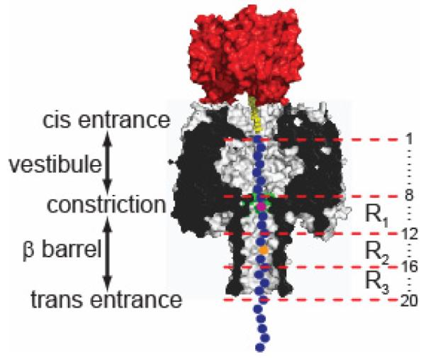
Schematic representation of an oligonucleotide (blue circles) immobilized inside an αHL pore (grey, cross-section) by the use of a 3′ biotin (yellow)•streptavidin (red) linkage (Figure S1). The bases are numbered (right) relative to the 3′ biotinylated end of the DNA. The αHL pore can be divided into two halves, each approximately 5 nm in length: an upper cap domain located between the cis entrance and the constriction, containing a roughly spherical vestibule, and a fourteen-stranded, transmembrane, antiparallel β barrel, located between the constriction and the trans entrance. The constriction of 1.4 nm diameter is formed by the Glu-111, Met-113 and Lys-147 (all three shaded green) side chains contributed by all seven subunits. R1, R2 and R3 represent the three base recognition sites in the αHL nanopore within the β barrel domain of the pore.
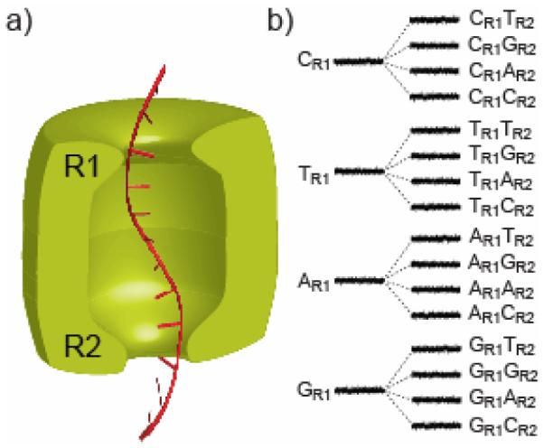
a) A hypothetical nanopore sensor (green) with two reading heads, R1 and R2, which could in principle extract more sequence information from a DNA strand (red) than a device with a single reading head. b) To illustrate the idea, we assume that the four bases of DNA at reading head R1 produce 4 distinct current levels (widely dispersed as shown). Each of the levels is split into 4 additional levels (with a lesser dispersion, for the purpose of illustration) by the second reading head R2, yielding 16 current levels in total and providing redundant information about the DNA sequence.
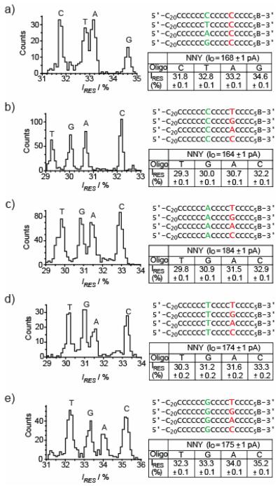
Histograms of residual current levels for E111N/K147N/M113Y (NNY) pores are shown (left), for a set of 4 oligonucleotides (right). B represents the 3′ biotin-TEG extension (Figure S1). Each experiment was conducted at least three times, and the results displayed in the figure are from a single experiment. When the oligonucleotides are driven into the αHL pore the substituted nucleotides are positioned at R1 (red) or R2 (green). Gaussian fits were performed for each peak in the histograms and the mean value of the residual current (IRES) for each oligonucleotide is displayed in the tables to the right of the histograms and in Tables S1-5 for panels a-e respectively.
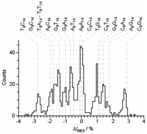
E111N/K147N/M113Y (NNY) pores were probed with 16 oligonucleotides, with the sequence 5′–CCCCCCCCCCCCCCCCCCCCCCCCCCNCCCCNCCCCCCCCB–3′, where N is A, T, G or C (N9N14, Table S8). B represents the 3′ biotin-TEG extension (Figure S1). A histogram displaying the residual current level differences (Table S9) for blockades by the various oligonucleotides, relative to the mean blockade produced by poly(dC) is shown. The current level for poly(dC) is set as zero. Blockades which have a residual current level lower than poly(dC) have negative ΔIRES values and blockades which have higher residual current levels than poly(dC) have positive ΔIRES values. The grey dashed lines show the predicted residual current levels, based on the ΔIRES data displayed in Table S6 (see the text). The predicted and measured ΔIRES values are displayed in Table S7.
References
-
- Branton D, Deamer DW, Marziali A, Bayley H, Benner SA, Butler T, Di Ventra M, Garaj S, Hibbs A, Huang X, Jovanovich SB, Krstic PS, Lindsay S, Ling XS, Mastrangelo CH, Meller A, Oliver JS, Pershin YV, Ramsey JM, Riehn R, Soni GV, Tabard-Cossa V, Wanunu M, Wiggin M, Schloss JA. Nature Biotechnology. 2008;26:1146. - PMC - PubMed
-
- Purnell RF, Schmidt JJ. ACS Nano. 2009 - PubMed
Publication types
MeSH terms
Substances
Grants and funding
LinkOut - more resources
Full Text Sources
Other Literature Sources
