. 2010 Jan;48(1):20-30.
doi: 10.1002/dvg.20578.

The zebrafish dyrk1b gene is important for endoderm formation

Affiliations

The zebrafish dyrk1b gene is important for endoderm formation

Gohar Mazmanian et al. Genesis. 2010 Jan.

Abstract

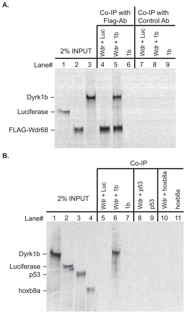
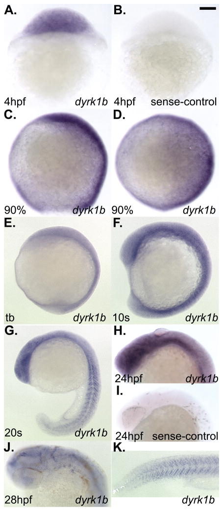
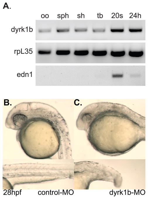
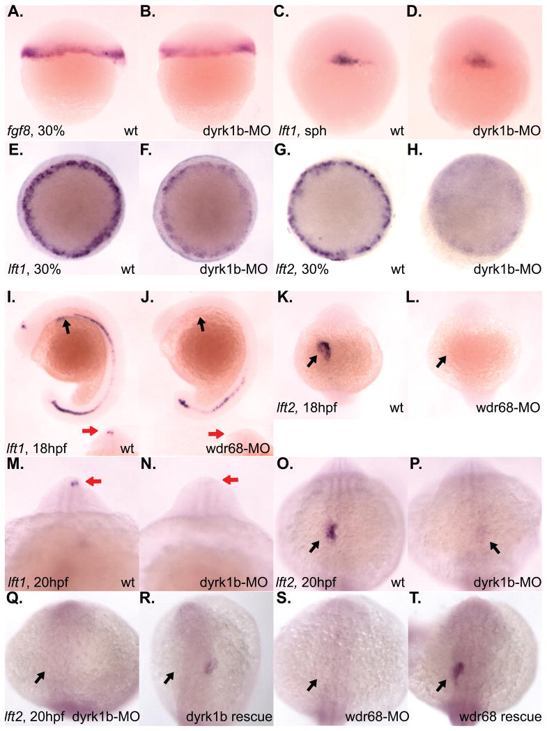
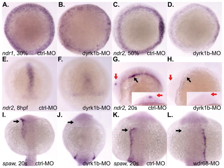
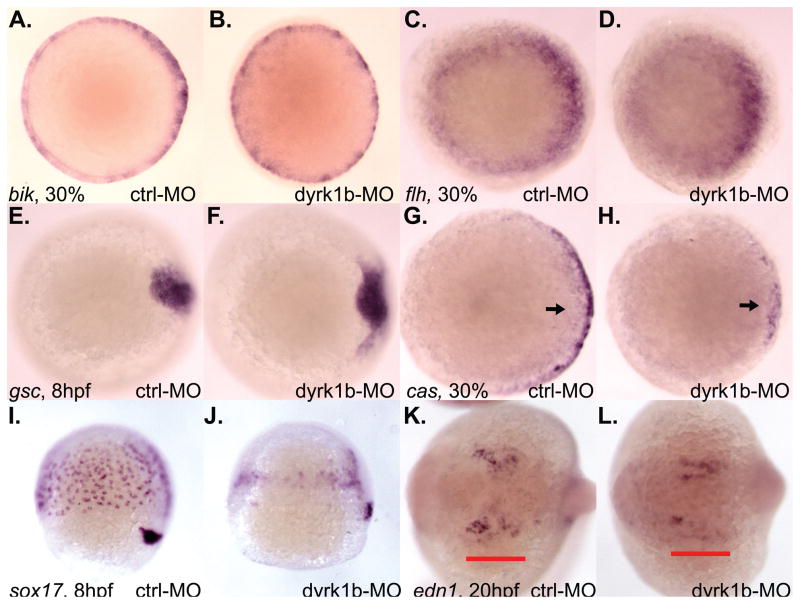
Nodal-signaling is required for specification of mesoderm, endoderm, establishing left-right asymmetry, and craniofacial development. Wdr68 is a WD40-repeat domain-containing protein recently shown to be required for endothelin-1 (edn1) expression and subsequent lower jaw development. Previous reports detected the Wdr68 protein in multiprotein complexes containing mammalian members of the dual-specificity tyrosine-regulated kinase (dyrk) family. Here we describe the characterization of the zebrafish dyrk1b homolog. We report the detection of a physical interaction between Dyrk1b and Wdr68. We also found perturbations of nodal signaling in dyrk1b antisense morpholino knockdown (dyrk1b-MO) animals. Specifically, we found reduced expression of lft1 and lft2 (lft1/2) during gastrulation and a near complete loss of the later asymmetric lft1/2 expression domains. Although wdr68-MO animals did not display lft1/2 expression defects during gastrulation, they displayed a near complete loss of the later asymmetric lft1/2 expression domains. While expression of ndr1 was not substantially effected during gastrulation, ndr2 expression was moderately reduced in dyrk1b-MO animals. Analysis of additional downstream components of the nodal signaling pathway in dyrk1b-MO animals revealed modestly expanded expression of the dorsal axial mesoderm marker gsc while the pan-mesodermal marker bik was largely unaffected. The endodermal markers cas and sox17 were also moderately reduced in dyrk1b-MO animals. Notably, and similar to defects previously reported for wdr68 mutant animals, we also found reduced expression of the pharyngeal pouch marker edn1 in dyrk1b-MO animals. Taken together, these data reveal a role for dyrk1b in endoderm formation and craniofacial patterning in the zebrafish.


Figures

Figure 1
Figure 1. The zebrafish Wdr68 and Dyrk1b proteins can physically interact
A) Lane 1 shows 2% of the negative control Luciferase protein input. Lane 2 shows 2% of the FLAG-Wdr68 protein input. Lane 3 shows 2% of the Dyrk1b protein input. Lane 4 shows the results of a co-immunoprecipitation (co-IP) between FLAG-Wdr68 and Luciferase indicating no co-IP of the negative control Luciferase. Lane 5 shows the results of a co-IP between FLAG-Wdr68 and Dyrk1b indicating that Dyrk1b can physically interact with FLAG-Wdr68. Lane 6 shows the dependence of the Dyrk1b co-IP on the presence of FLAG-Wdr68. Lanes 7–9 are the same as lanes 4–6 but with the FLAG antibody substituted with an unrelated histone H3 control antibody. B) Lane 1 shows 2% of Dyrk1b input. Lane 2 shows 2% of negative control Luciferase input. Lane 3 shows 2% of negative control p53 input. Lane 4 shows 2% of negative control hoxb8a input. The FLAG-Wdr68 protein used in these experiments was translated with non-radioactive amino acids and is therefore not detected in the image. Lane 5 shows the results of a co-IP between FLAG-Wdr68 and Luciferase indicating no co-IP of the negative control Luciferase. Lane 6 shows the results of a co-IP between FLAG-Wdr68 and Dyrk1b indicating that Dyrk1b can physically interact with FLAG-Wdr68. Lane 7 shows the dependence of the Dyrk1b co-IP on the presence of FLAG-Wdr68. Lanes 8–9 show the lack of interaction between FLAG-Wdr68 and the negative control p53. Lanes 10–11 show the lack of interaction between FLAG-Wdr68 and the negative control hoxb8a.
Figure 2
Figure 2. Whole-mount in situ hybridization analysis of dyrk1b expression in wildtype zebrafish embryos
A) ubiquitous expression of dyrk1b at 4hpf. B) sense-strand negative control for staining also at 4hpf. C) lateral view of 90% epiboly stage, D) dorsal view of 90% epiboly stage. E) lateral view of tailbud (tb) stage. F) lateral views of 10 somites (10s) stage. G) lateral view of 20 somites (20s) stage. H) lateral view of 24hpf stage. I) sense-strand negative control for staining also at 24hpf. J) dorsolateral view of 28hpf stage. K) lateral tail view of the same animal shown in panel J. Scale bar in panel B = 100 micrometers length.
Figure 3
Figure 3. dyrk1b is maternally supplied and essential during embryonic development in the zebrafish
A) RT-PCR detection of dyrk1b transcripts in unfertilized oocytes (oo), sphere stage (sph), shield stage (sh), tailbud stage (tb), 20 somites stage (20s), and 24 hours post fertilization (24h) stage animals. The rpl35 gene serves as positive control. The edn1 gene serves as negative control for detection in oocytes. B) Normal phenotype of 5-mismatch control morpholino-injected (control-MO) animals at 28hpf. Upper panel shows normal head, eye, pigment and ear development. Lower panel shows normal notochord, tail and somite development. C) Phenotype of dyrk1b antisense morpholino-injected (dyrk1b-MO) animals at 28hpf. Upper panel shows small head and eyes of the morphant animals. Lower panel shows the moderately shortened length of the tail.
Figure 4
Figure 4. Reduced expression of lft1 and lft2 in dyrk1b and wdr68 knockdown animals
A) lateral view of normal fgf8 expression in control animals at 30% epiboly stage. B) normal fgf8 expression in dyrk1b-MO animals at 30% epiboly stage. C) lateral view of normal lft1 expression in control animals at sphere (sph) stage. D) normal lft1 expression in dyrk1b-MO animals at sphere stage. E) dorsal view of normal lft1 expression in control animals at 30% epiboly stage. F) reduced lft1 expression in dyrk1b-MO animals at 30% epiboly stage. G) dorsal view of normal lft2 expression in control animals at 30% epiboly stage. H) reduced lft2 expression in dyrk1b-MO animals at 30% epiboly stage. I) lateral view of normal lft1 expression in control animals at 18hfp. Black arrows indicate asymmetric expression in the lateral plate mesoderm of the developing heart field. Red arrows in inset images are on the left side of the animals and indicate asymmetric expression in the diencephalon. J) severely reduced lft1 expression in the asymmetric heart and diencephalon territories of wdr68-MO animals at 18hpf. K) dorsal view of normal lft2 expression in control animals at 18hpf. Black arrows indicate asymmetric expression in the lateral plate mesoderm of the developing heart field. L) severely reduced lft2 expression in the asymmetric heart and diencephalon territories of wdr68-MO animals at 18hpf. M) anterior view of normal lft1 expression in the asymmetric diencephalon territory of control animals at 20hpf. Red arrows are on the left side of the animals and indicate asymmetric expression in the diencephalon. N) severely reduced lft1 expression in the asymmetric diencephalon territory of dyrk1b-MO animals at 20hpf. O) dorsal view of normal lft2 expression in the asymmetric heart territory of control animals at 20hpf. Black arrows indicate asymmetric expression in the lateral plate mesoderm of the developing heart field. P) severely reduced lft2 expression in the asymmetric heart territory of dyrk1b-MO animals at 20hpf. Q) dorsal view of severely reduced lft2 expression in dyrk1b-MO animals co-injected with EF1alpha transcripts. R) partial rescue of lft2 expression in dyrk1b-MO animals co-injected with Dyrk1b transcripts. S) dorsal view of severely reduced lft2 expression in wdr68-MO animals co-injected with EF1alpha transcripts. T) partial rescue of lft2 expression in wdr68-MO animals co-injected with FLAG-Wdr68 transcripts.
Figure 5
Figure 5. dyrk1b and wdr68 are important for ndr2 and spaw expression in the zebrafish
A) dorsal view of normal ndr1 expression in control animals at 30% epiboly stage. B) normal ndr1 expression in dyrk1b-MO animals at 30% epiboly stage. C) A) normal ndr2 expression in control animals at 50% epiboly stage. D) reduced ndr2 expression in dyrk1b-MO animals at 50% epiboly stage. E) shield view of normal ndr2 expression in control animals at 8hpf. F) reduced ndr2 expression in dyrk1b-MO animals at 8hpf. G) normal asymmetric ndr2 expression in control animals at 20 somites stage. Black arrow indicates asymmetric expression in lateral plate mesoderm. Red arrows indicate asymmetric expression in diencephalon. Red arrow in inset image is on left side of animal. H) reduced ndr2 expression in dyrk1b-MO animals at 20 somites stage. I) dorso-posterior view of normal asymmetric spaw expression in control animals at 20 somites stage. J) reduced expression of spaw in dyrk1b-MO animals at 20 somites stage. K) dorsal view of normal asymmetric spaw expression in control animals at 20 somites stage. L) loss of asymmetry in the expression of spaw in wdr68-MO animals at 20 somites stage.
Figure 6
Figure 6. dyrk1b is important for endoderm induction and edn1 expression in the zebrafish
A) normal bik expression in control animals at 30% epiboly stage. B) normal bik expression in dyrk1b-MO animals at 30% epiboly stage. C) normal flh expression in control animals at 30% epiboly stage. D) normal flh expression in dyrk1b-MO animals at 30% epiboly stage. E) normal gsc expression in control animals at 8hpf. F) modest expansion of gsc expression in dyrk1b-MO animals at 8hpf. G) normal cas expression in control animals at 30% epiboly stage. H) reduced cas expression in dyrk1b-MO animals at 30% epiboly stage. I) normal sox17 expression in control animals at 8hpf. J) reduced sox17 expression in dyrk1b-MO animals at 8hpf. K) normal edn1 expression in control animals at 20hpf. Red bar underlines pharyngeal expression domain of edn1. L) reduced edn1 expression in dyrk1b-MO animals at 20hpf.

Similar articles

Cited by

References

    1. Alexander J, Stainier DY. A molecular pathway leading to endoderm formation in zebrafish. Curr Biol. 1999;9:1147–1157. - PubMed
    1. Amsterdam A, Nissen RM, Sun Z, Swindell EC, Farrington S, Hopkins N. Identification of 315 genes essential for early zebrafish development. Proc Natl Acad Sci U S A. 2004;101:12792–12797. - PMC - PubMed
    1. Chen Y, Schier AF. Lefty proteins are long-range inhibitors of squint-mediated nodal signaling. Curr Biol. 2002;12:2124–2128. - PubMed
    1. Clouthier DE, Hosoda K, Richardson JA, Williams SC, Yanagisawa H, Kuwaki T, Kumada M, Hammer RE, Yanagisawa M. Cranial and cardiac neural crest defects in endothelin-A receptor-deficient mice. Development. 1998;125:813–824. - PubMed
    1. Clouthier DE, Schilling TF. Understanding endothelin-1 function during craniofacial development in the mouse and zebrafish. Birth Defects Res C Embryo Today. 2004;72:190–199. - PubMed

Publication types

MeSH terms