

Complete response to radiation therapy of orbital metastasis from hepatocellular carcinoma
- PMID: 20014466
- PMCID: PMC2795189
- DOI: 10.3748/wjg.15.6000
Complete response to radiation therapy of orbital metastasis from hepatocellular carcinoma
Abstract
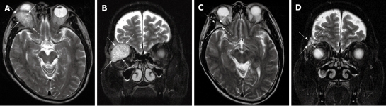
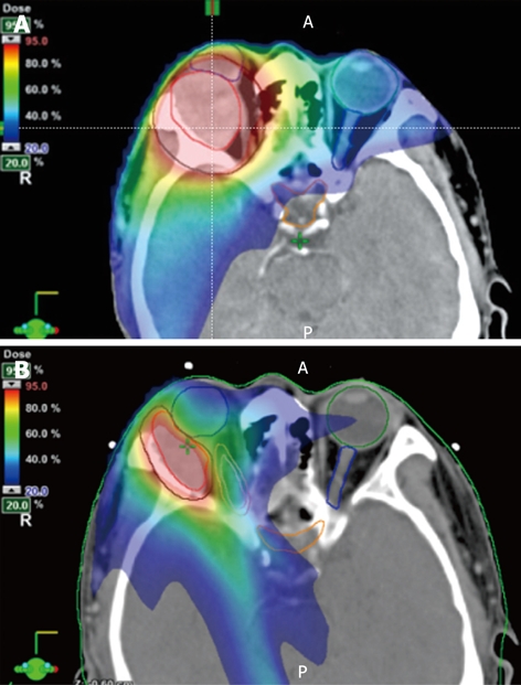
The incidence of hepatocellular carcinoma (HCC) is increasing in the United States, and 50%-75% of patients with HCC will develop metastatic disease. Orbital metastases from HCC are extremely rare. We report the case of a 52-year-old male with known metastatic HCC, who presented with severe proptosis and diplopia. An orbital mass was identified on magnetic resonance imaging (MRI) and confirmed to have hypermetabolic activity on positron emission tomography/computed tomography. He received a palliative course of external beam radiation therapy to the right orbit. Intensity modulated radiation therapy (IMRT) was used to allow sparing of critical normal tissues in close proximity to the tumor. One month after completion of IMRT to 58 Gray in 30 fractions delivered over 6 wk, the patient had a complete clinical, radiologic (MRI) and symptomatic response. The patient continues to have local control in the orbit 1.7 years after therapy completion. All critical normal structures were kept below the tolerance dose using IMRT, and no toxicities were observed.
Keywords: Eye neoplasms; Hepatocellular carcinoma; Intensity modulated radiation therapy; Metastasis; Palliative therapy.
Figures
Similar articles
-
Cytologic diagnosis of metastatic hepatocellular carcinoma presenting as an orbital mass. A case report.Acta Cytol. 2007 Jan-Feb;51(1):83-5. doi: 10.1159/000325689. Acta Cytol. 2007. PMID: 17328502
-
Hepatocellular carcinoma metastatic to the orbit: a case report.Tokai J Exp Clin Med. 2006 Apr 20;31(1):7-10. Tokai J Exp Clin Med. 2006. PMID: 21302214
-
Hepatocellular carcinoma metastatic to the orbit.Arch Ophthalmol. 1998 Jul;116(7):942-5. doi: 10.1001/archopht.116.7.942. Arch Ophthalmol. 1998. PMID: 9682712 Review.
-
Orbital metastasis of hepatocellular carcinoma: a case report.West Afr J Med. 2011 Jul-Aug;30(4):305-7. West Afr J Med. 2011. PMID: 22669839
-
Orbital metastasis from hepatocellular carcinoma.Surv Ophthalmol. 2005 Sep-Oct;50(5):485-9. doi: 10.1016/j.survophthal.2005.06.014. Surv Ophthalmol. 2005. PMID: 16139041 Review.
Cited by
-
Hepatocellular Carcinoma with Orbital Metastasis: a Unique Multidisciplinary Case Report.J Gastrointest Cancer. 2019 Dec;50(4):978-982. doi: 10.1007/s12029-018-0159-3. J Gastrointest Cancer. 2019. PMID: 30105522 No abstract available.
-
Ocular Metastatic Tumor in a Patient With Hepatocellular Carcinoma.Cureus. 2024 Nov 25;16(11):e74435. doi: 10.7759/cureus.74435. eCollection 2024 Nov. Cureus. 2024. PMID: 39723270 Free PMC article.
-
Radiotherapy of orbital metastases: a systematic review of management and treatment outcomes on behalf of palliative care study group of Italian association of radiotherapy and clinical oncology (AIRO).Br J Radiol. 2023 Nov;96(1151):20230124. doi: 10.1259/bjr.20230124. Epub 2023 Oct 11. Br J Radiol. 2023. PMID: 37751164 Free PMC article.
-
Orbital metastasis from hepatocellular carcinoma revealed by sudden exophthalmos.BMJ Case Rep. 2021 May 12;14(5):e242136. doi: 10.1136/bcr-2021-242136. BMJ Case Rep. 2021. PMID: 33980563 Free PMC article.
-
Prediction model of ocular metastasis from primary liver cancer: Machine learning-based development and interpretation study.Cancer Med. 2023 Oct;12(20):20482-20496. doi: 10.1002/cam4.6540. Epub 2023 Oct 5. Cancer Med. 2023. PMID: 37795569 Free PMC article.
References
-
- Ries LAG, Melbert D, Krapcho M, Stinchcomb DG, Howlader N, Horner MJ, Mariotto A, Miller BA, Feuer EJ, Altekruse SF, Lewis DR, Clegg L, Eisner MP, Reichman M, Edwards BK, editors. SEER Cancer Statistics Review, 1975-2005, National Cancer Institute. Bethesda, MD, based on November 2007 SEER data submission, posted to the SEER web site, 2008. Available from: http://seer.cancer.gov/csr/1975_2005/
-
- El-Serag HB. Hepatocellular carcinoma: recent trends in the United States. Gastroenterology. 2004;127:S27–S34. - PubMed
-
- Parkin DM, Bray F, Ferlay J, Pisani P. Global cancer statistics, 2002. CA Cancer J Clin. 2005;55:74–108. - PubMed
-
- Srinivasan R, Krishnanand G. Cytologic diagnosis of metastatic hepatocellular carcinoma presenting as an orbital mass. A case report. Acta Cytol. 2007;51:83–85. - PubMed
-
- Zubler MA, Rivera R, Lane M. Hepatoma presenting as a retro-orbital metastasis. Cancer. 1981;48:1883–1885. - PubMed
Publication types
MeSH terms
Grants and funding
LinkOut - more resources
Full Text Sources
Medical
