

doi: 10.1021/ja908467y.
Optical imaging of mammary and prostate tumors in living animals using a synthetic near infrared zinc(II)-dipicolylamine probe for anionic cell surfaces
Affiliations
- PMID: 20014845
- PMCID: PMC2805267
- DOI: 10.1021/ja908467y
Item in Clipboard
Optical imaging of mammary and prostate tumors in living animals using a synthetic near infrared zinc(II)-dipicolylamine probe for anionic cell surfaces
J Am Chem Soc.
.
Abstract
In vivo optical imaging shows that a fluorescent imaging probe, comprised of a near-infrared fluorophore attached to an affinity group containing two zinc(II)-dipicolylamine (Zn-DPA) units, targets prostate and mammary tumors in two different xenograft animal models. The tumor selectivity is absent with control fluorophores whose structures do not have appended Zn-DPA targeting ligands. Ex vivo biodistribution and histological analyses indicate that the probe is targeting the necrotic regions of the tumors, which is consistent with in vitro microscopy showing selective targeting of the anionic membrane surfaces of dead and dying cells.
Figures
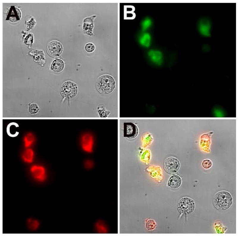
Micrographs (60X magnification) of Jurkat cells treated with cytotoxic camptothecin (10 μM) for 3.5 h and stained simultaneously with Annexin V-Alexa Fluor 488, and probe 1 (10 μM). Brightfield image of the entire field of cells (A); cells stained with Annexin V-Alexa Fluor 488 (B); cells stained with probe 1 (C); overlay of images A, B, and C (D). No staining of healthy cells was observed in the absence of camptothecin.
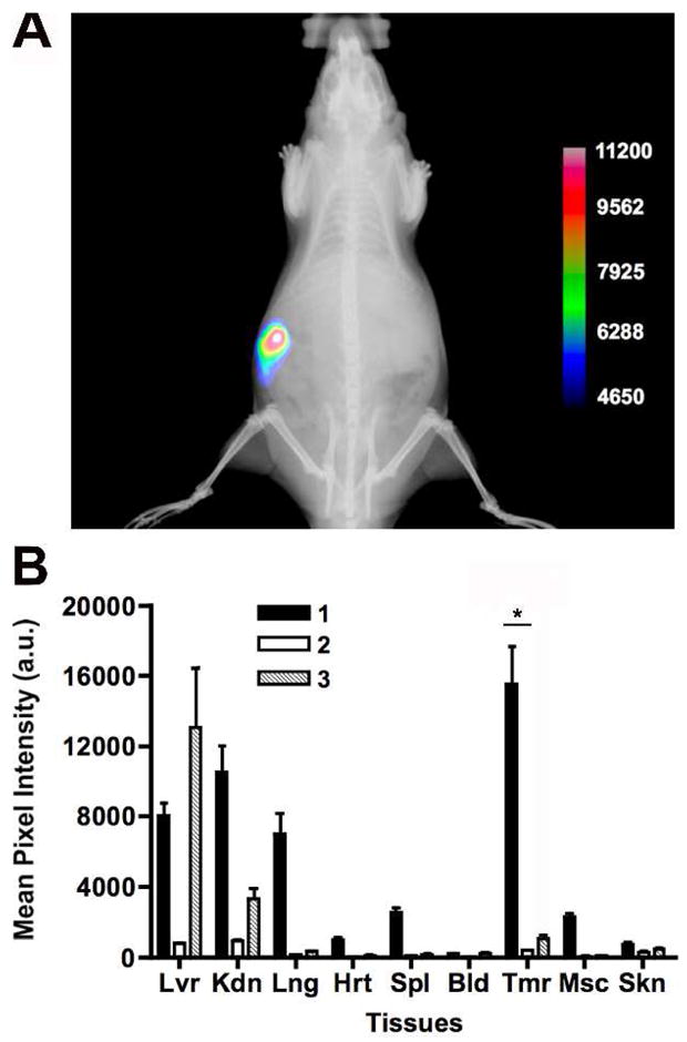
X-ray and fluorescence overlay image of a rat prostate tumor model at 24 h post-injection of probe 1 (A). The image was acquired at a 190 mm field of view. Bar graph showing ex vivo tissue distribution of probes 1, 2, and 3 (B). The values represent the mean (n=3), ± standard error of the mean. *P<0.0005. This imaging data is representative of four replicate studies using independent cohorts.
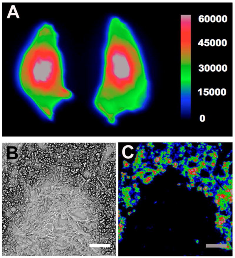
Ex vivo analysis of probe 1 localization in rat prostate tumor. Excised PAIII prostate tumors were sliced along the longest axis, and a 30 mm field of view generated the representative near-IR fluorescence intensity image (A). Representative co-registered micrographs of a 5 μm histological slice of tumor core; the brightfield image (B) shows necrotic cells as darker regions that colocalize with near-IR fluorescence intensity image from probe 1 (C). Scale bar = 100 μm.
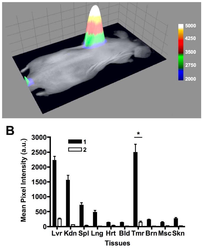
Representative overlay image of a nude mouse with an EMT-6 mammary tumor. Brightfield and fluorescence intensity images were acquired 24 hrs following injection of probe 1 (A). The fluorescence intensity is plotted along the z-axis of this 3D surface diagram. Images were taken at an 80 mm field of view. Bar graph showing ex vivo tissue distribution of probe 1 and control 2 (B). The values represent the mean (n=3), ± standard error of the mean. *P<0.005. This imaging data is representative of three replicate studies using independent cohorts.
Similar articles
-
Deep-red fluorescent imaging probe for bacteria.Bioorg Med Chem Lett. 2012 Apr 15;22(8):2833-6. doi: 10.1016/j.bmcl.2012.02.078. Epub 2012 Mar 3. Bioorg Med Chem Lett. 2012. PMID: 22424976 Free PMC article.
-
Enhanced cell death imaging using multivalent zinc(II)-bis(dipicolylamine) fluorescent probes.Mol Pharm. 2013 Sep 3;10(9):3296-303. doi: 10.1021/mp300720k. Epub 2013 Aug 5. Mol Pharm. 2013. PMID: 23915311 Free PMC article.
-
Phenoxide-Bridged Zinc(II)-Bis(dipicolylamine) Probes for Molecular Imaging of Cell Death.Bioconjug Chem. 2016 Feb 17;27(2):363-75. doi: 10.1021/acs.bioconjchem.5b00447. Epub 2015 Sep 11. Bioconjug Chem. 2016. PMID: 26334386 Free PMC article.
-
Library synthesis, screening, and discovery of modified Zinc(II)-Bis(dipicolylamine) probe for enhanced molecular imaging of cell death.Bioconjug Chem. 2014 Apr 16;25(4):724-37. doi: 10.1021/bc500003x. Epub 2014 Mar 13. Bioconjug Chem. 2014. PMID: 24575875 Free PMC article.
-
Anion recognition and sensing with Zn(II)-dipicolylamine complexes.Chem Soc Rev. 2012 Jul 21;41(14):4928-65. doi: 10.1039/c2cs35087d. Epub 2012 Jun 12. Chem Soc Rev. 2012. PMID: 22688834 Review.
Cited by
-
Semi-automatic synthesis and biodistribution of N-(2-18F-fluoropropionyl)-bis(zinc (II)-dipicolylamine) (18F-FP-DPAZn2) for AD model imaging.BMC Med Imaging. 2017 Apr 21;17(1):27. doi: 10.1186/s12880-017-0200-1. BMC Med Imaging. 2017. PMID: 28431519 Free PMC article.
-
Near Infrared Fluorescence Imaging in Nano-Therapeutics and Photo-Thermal Evaluation.Int J Mol Sci. 2017 Apr 28;18(5):924. doi: 10.3390/ijms18050924. Int J Mol Sci. 2017. PMID: 28452928 Free PMC article. Review.
-
Selective non-covalent triggered release from liposomes.Chem Commun (Camb). 2012 Aug 21;48(65):8123-5. doi: 10.1039/c2cc32962j. Epub 2012 Jul 9. Chem Commun (Camb). 2012. PMID: 22772732 Free PMC article.
-
A nanoparticle formula for delivering siRNA or miRNAs to tumor cells in cell culture and in vivo.Nat Protoc. 2014 Aug;9(8):1900-15. doi: 10.1038/nprot.2014.128. Epub 2014 Jul 17. Nat Protoc. 2014. PMID: 25033207 Free PMC article.
-
Avenues to molecular imaging of dying cells: Focus on cancer.Med Res Rev. 2018 Sep;38(6):1713-1768. doi: 10.1002/med.21495. Epub 2018 Mar 12. Med Res Rev. 2018. PMID: 29528513 Free PMC article. Review.
References
-
- Kulasingam V, Diamandis EP. Nat Clin Pract Oncol. 2008;5:588–599. - PubMed
-
- Elliot JI, Surprenant A, Marelli-Berg FM, Cooper JC, Cassady-Cain RL, Wooding C, Linton K, Alexander DR, Higgins CF. Nat Cell Biol. 2005;8:808–816. - PubMed
-
- Ran S, Downes A, Thorpe PA. Cancer Res. 2002;62:6132–6140. - PubMed
-
- Distler JHW, Huber LC, Reich CF, III, Gay S, Distler O, Pisetsky DS. Apoptosis. 2005;10:731–741. - PubMed
Publication types
MeSH terms
Substances
Grants and funding
LinkOut - more resources
Full Text Sources
Other Literature Sources
Medical
