

Review
doi: 10.1021/cb900256m.
Making the cut: the chemical biology of cytokinesis
Affiliations
- PMID: 20014865
- PMCID: PMC2807474
- DOI: 10.1021/cb900256m
Item in Clipboard
Review
Making the cut: the chemical biology of cytokinesis
ACS Chem Biol.
.
Abstract
Cytokinesis is the last step in the cell cycle, where daughter cells finally separate. It is precisely regulated in both time and space to ensure that each daughter cell receives an equal share of DNA and other cellular materials. Chemical biology approaches have been used very successfully to study the mechanism of cytokinesis. In this review, we discuss the use of small molecule probes to perturb cytokinesis, as well as the role naturally occurring small molecule metabolites such as lipids play during cytokinesis.
Figures
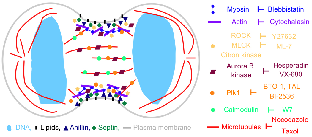
During cytokinesis, a contractile ring assembles. It is located just beneath the plasma membrane and includes a network of actin and myosin II filaments as well as septins and Anillin. Phosphorylation and activation of myosin II at the cleavage furrow is regulated by several kinases, including ROCK, MLCK and Citron kinase. The mitotic kinases Plk1 and Aurora B both localize to the cleavage furrow as well as midzone microtubules where they function as key regulators of cytokinesis. An expanding collection of small molecules has been used to dissect cytokinesis regulation in both time and space. Key cytokinesis protein and their corresponding small molecule inhibitors are shown. For clarity, only localizations at the cleavage furrow and midzone microtubules are shown. Not drawn to scale.
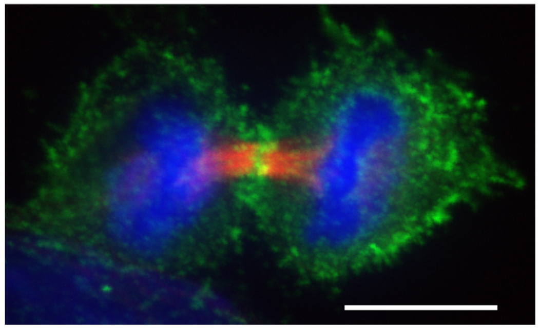
Phosphorylated myosin II accumulates at the cleavage furrow.  (red) were stained with anti-tubulin (Sigma T6199),
  (green) was stained with Phospho-Myosin Light Chain 2 (Ser19) antibody (Cell Signalling, 3671L),
  (blue) with DAPI. Scalebar represents 10 µM.
(red) were stained with anti-tubulin (Sigma T6199), (green) was stained with Phospho-Myosin Light Chain 2 (Ser19) antibody (Cell Signalling, 3671L), (blue) with DAPI. Scalebar represents 10 µM.
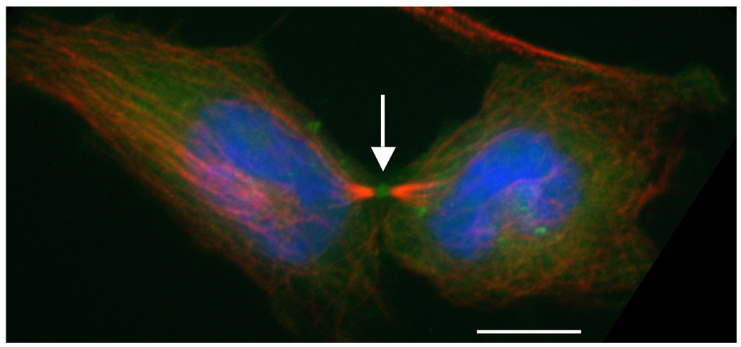
PIP2 localizes to the midbody (white arrow). HeLa cells were transfected with  (vector kindly provided by Tamas Balla, NIH). (red) were stained with anti-tubulin (Sigma T6199), (blue) with DAPI. Scalebar represents 10 µM.
(vector kindly provided by Tamas Balla, NIH). (red) were stained with anti-tubulin (Sigma T6199), (blue) with DAPI. Scalebar represents 10 µM.References
-
- Eggert US, Mitchison TJ, Field CM. ANIMAL CYTOKINESIS: From Parts List to Mechanisms. Annu Rev Biochem. 2006;75:543–566. - PubMed
-
- D'Avino PP, Savoian MS, Glover DM. Cleavage furrow formation and ingression during animal cytokinesis: a microtubule legacy. J Cell Sci. 2005;118:1549–1558. - PubMed
-
- Glotzer M. The molecular requirements for cytokinesis. Science. 2005;307:1735–1739. - PubMed
-
- Maddox AS, Oegema K. Deconstructing cytokinesis. Nat Cell Biol. 2003;5:773–776. - PubMed
Publication types
MeSH terms
Grants and funding
LinkOut - more resources
Full Text Sources
