Review
. 2010 Feb;43(1):76-83.
doi: 10.3109/08916930903374618.

Regulation of autoreactive B cell responses to endogenous TLR ligands

Affiliations
Review

Regulation of autoreactive B cell responses to endogenous TLR ligands

Ana Maria Avalos et al. Autoimmunity. 2010 Feb.

Abstract

Immune complexes containing DNA and RNA are responsible for disease manifestations found in patients with systemic lupus erythematosus (SLE). B cells contribute to SLE pathology through BCR recognition of endogenous DNA- and RNA- associated autoantigens and delivery of these self-constituents to endosomal TLR9 and TLR7, respectively. B cell activation by these pathways leads to production of class-switched DNA- and RNA-reactive autoantibodies, contributing to an inflammatory amplification loop characteristic of disease. Intriguingly, self-DNA and RNA are typically non-stimulatory for TLR9/7 due to the absence of stimulatory sequences or the presence of molecular modifications. Recent evidence from our laboratory and others suggests that B cell activation by BCR/TLR pathways is tightly regulated by surface-expressed receptors on B cells, and the outcome of activation depends on the balance of stimulatory and inhibitory signals. Either IFNalpha engagement of the type I IFN receptor or loss of IgG ligation of the inhibitory FcgammaRIIB receptor promotes B cell activation by weakly stimulatory DNA and RNA TLR ligands. In this context, autoreactive B cells can respond robustly to common autoantigens. These findings have important implications for the role of B cells in vivo in the pathology of SLE.


Figures

Figure 1
Figure 1
Immune complexes (ICs) incorporating a TLR7 or TLR9 ligand induce AM14 B cell proliferation. IgG2a anti-TNP antibodies in complex with TNP-conjugated BSA are recognized by the AM14 BCR (anti-IgG2a) and internalized, but do not promote AM14 B cell proliferation due to failure to engage TLR7 or 9 (left panel). Spontaneous ICs comprised of IgG2a anti-DNA or anti-chromatin in complex with endogenous DNA or CG-rich DNA fragments promote AM14 B cell proliferation through engagement of TLR9 (middle panel). AM14 B cells proliferate in a TLR7-dependent manner when anti-RNA IgG2a antibodies are complexed with endogenous RNA, in the presence of IFNα (right panel).
Figure 2
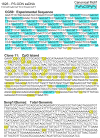
Figure 2. CpG-motif and CG-rich containing DNA fragments activate TLR9
The canonical CpG motif recognized by TLR9, PuPuCGPyPy is depicted in turquoise. The strong B cell mitogen 1826, a single stranded phosphorothioate (PS-DNA) oligonucleotide (ODN), contains two repeats of the CpG motif. CG50 is an experimentally designed phosphorodiester dsDNA fragment that includes 50 repeats of the canonical CpG motif. Clone 11 is a dsDNA fragment corresponding to an endogenous CpG island that contains two CpG motifs (in turquoise) and 42 CG-repeats (in yellow). SenP1 is a DNA fragment representative of genomic DNA.
Figure 3
Figure 3. The AM14 B cell response to ICs reflects the combined signal strength of the BCR, TLR7/9, IFNAR and FcγRIIB
(a) ICs that incorporate CG-poor DNA weakly engage the BCR (thin red arrow) and TLR9 (thin black arrow) and activation is prevented by weak engagement of FcγRIIB by IgG2a (thin blue arrow). This equilibrium can be disrupted by the addition of type I IFNs (green arrow), or loss of FcγRIIB engagement, resulting in B cell proliferation. (b) ICs that incorporate CG-rich DNA weakly engage the BCR and strongly engage TLR9 (thick black arrow) and activation cannot be prevented by weak engagement of FcγRIIB by IgG2a. (c) ICs that incorporate RNA weakly engage the BCR and TLR7 (thin purple arrow), and activation is prevented by weak engagement of FcγRIIB by IgG2a. This equilibrium can be disrupted by the addition of type I IFNs, or loss of FcγRIIB engagement, resulting in B cell proliferation. (d) ICs that incorporate CG-rich DNA weakly engage the BCR and robustly engage TLR9, and activation is suppressed by strong engagement of FcγRIIB by IgG2b (thick blue arrow). This response is amplified by loss of FcγRIIB engagement.

References

    1. Wolfowicz CB, Lyons S, Mahon CA, Marshak-Rothstein A, Rothstein TL. Isolation of self-recognizing IgG2a monoclonal rheumatoid factors. Clin Immunol Immunopathol. 1989;52:313–22. - PubMed
    1. Hannum LG, Ni D, Haberman AM, Weigert MG, Shlomchik MJ. A disease-related rheumatoid factor autoantibody is not tolerized in a normal mouse: implications for the origins of autoantibodies in autoimmune disease. J Exp Med. 1996;184:1269–78. - PMC - PubMed
    1. Wang H, Shlomchik MJ. Autoantigen-specific B cell activation in Fas-deficient rheumatoid factor immunoglobulin transgenic mice. J Exp Med. 1999;190:639–49. - PMC - PubMed
    1. Leadbetter EA, Rifkin IR, Hohlbaum AM, Beaudette BC, Shlomchik MJ, Marshak-Rothstein A. Chromatin-IgG complexes activate B cells by dual engagement of IgM and Toll-like receptors. Nature. 2002;416:603–7. - PubMed
    1. Viglianti GA, Lau CM, Hanley TM, Miko BA, Shlomchik MJ, Marshak-Rothstein A. Activation of autoreactive B cells by CpG dsDNA. Immunity. 2003;19:837–47. - PubMed

LinkOut - more resources