. 2010 Feb;43(1):103-10.
doi: 10.3109/08916930903384591.

RAGE-independent autoreactive B cell activation in response to chromatin and HMGB1/DNA immune complexes

Affiliations

RAGE-independent autoreactive B cell activation in response to chromatin and HMGB1/DNA immune complexes

Ana M Avalos et al. Autoimmunity. 2010 Feb.

Abstract

Increasing evidence suggests that the excessive accumulation of apoptotic or necrotic cellular debris may contribute to the pathology of systemic autoimmune disease. HMGB1 is a nuclear DNA-associated protein, which can be released from dying cells thereby triggering inflammatory processes. We have previously shown that IgG2a-reactive B cell receptor (BCR) transgenic AM14 B cells proliferate in response to endogenous chromatin immune complexes (ICs), in the form of the anti-nucleosome antibody PL2-3 and cell debris, in a TLR9-dependent manner, and that these ICs contain HMGB1. Activation of AM14 B cells by these chromatin ICs was inhibited by a soluble form of the HMGB1 receptor, RAGE-Fc, suggesting HMGB1-RAGE interaction was important for this response. To further explore the role of HMGB1 in autoreactive B cell activation, we assessed the capacity of purified calf thymus HMGB1 to bind dsDNA fragments and found that HMGB1 bound both CG-rich and CG-poor DNA. However, HMGB1-DNA complexes could not activate AM14 B cells unless HMGB1 was bound by IgG2a and thereby able to engage the BCR. To ascertain the role of RAGE in autoreactive B cell responses to chromatin ICs, we intercrossed AM14 and RAGE-deficient mice. We found that spontaneous and defined DNA ICs activated RAGE+ and RAGE(- ) AM14 B cells to a comparable extent. These results suggest that HMGB1 promotes B cell responses to endogenous TLR9 ligands through a RAGE-independent mechanism.


Figures
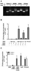
Figure 1
Figure 1. HMGB1 binds mammalian DNA in a sequence independent manner, and promotes TLR9-dependent responses when bound to CG-rich DNA and delivered through the BCR
(A) CG-rich (Clone 11 and CG50) and CG-poor (SUMO and CGneg) dsDNA at 800 ng/ml were incubated in the presence or absence of 10 μg/ml of HMGB1 for 1 h at 37°C, and ran in a agarose gel with Ethidium bromide. (B) 10 μg/ml of HMGB1 alone or in the presence of 250 ng/ml of unlabelled dsDNA fragments CG50 (G50) or CGneg (Gng) were added to AM14 B cells. HMGB1 alone or in the presence of CG50, CGneg, Clone 11 (C11) or Sumo (SU) were incubated before addition of 5 μg/ml of anti-HMGB1 IgG2a antibody. The resulting complexes were added to AM14 B cells and proliferation determined by [3H] thymidine incorporation. The TLR2 ligand Pam4CysK4 (P3Cysk) at 1 μg/ml was used as a control. (C) HMGB1 alone or in complex with anti-HMGB1, or CG50/HMGB1/anti-HMGB1 complexes were added to AM14 B cells from TLR9 sufficient (grey bars) or TLR9−/− mice (white bars). As controls, the TLR9 ligand 1826 (CpG) and TLR2 ligand Pam3CysK4 (P3CysK), both at 1 μg/ml were added. In (A), shown is a representative experiment of at least three. (B) and (C), shown are the mean+SEM of three independent experiments.
Figure 2
Figure 2. HMGB1 does not enhance AM14 B cell proliferation by DNA-fragment ICs
(A) AM14 B cells were incubated with PL2-3 (left) or PA4 (right) in the presence (open squares) or absence (closed circles) of 1 μg/ml HMGB1. (B) AM14 B cells were incubated with the anti-DNA IgG2a antibody PA4, PA4/CG50, PA4/CGneg, anti-biotin IgG2a 1D4, 1D4/Bio-Clone 11, 1D4/Bio-Sumo in the absence (black bars) or the presence (open bars) of 1μg/ml HMGB1. As control, HMGB1 was incubated in the presence of the TLR2 ligand Pam3CysK4. In (A) and (B), proliferation was determined as [3H] thymidine incorporation. Shown are a representative experiment (A) and mean+SEM of four experiments, except for 1D4/C11 and 1D4/SUMO, with mean+SEM of three (B).
Figure 3
Figure 3. RAGE deficiency does not affect B cell phenotype or responses to mitogens
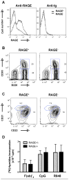
(A) Splenic naïve B cells from RAGE+/− (RAGE+) and RAGE−/− (RAGE) mice were stained with mouse anti-mouse RAGE antibody (left panel) or with mouse anti-Ig (right panel). (B and C) B220+ splenocytes from RAGE+ and RAGE mice were stained using anti-mouse CD93 (B) or anti-mouse CD21 and CD23 antibodies (C). Numbers represent percentages of each population. (D) Splenic naïve B cells from RAGE+ (grey bars) and RAGE (black bars) mice were incubated with 50 μg/ml anti-IgM F(ab)′2, 0.1 μg/ml of the TLR9 ligand CpG ODN 1826 (CpG) and 30 ng/ml of the TLR7 ligand R848, and proliferation determined by [3H] thymidine incorporation. In (AC), shown are representative results of three (A) and six (B and C) independent experiments. In (D), shown are mean+SEM of four (F(ab)′2), six (CpG) and three (R848) independent experiments.
Figure 4
Figure 4. RAGE-deficient AM14 B cells respond to chromatin ICs comparably to RAGE sufficient cells
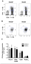
(A and B) B220+ splenocytes from AM14 RAGE+ and RAGE mice were stained using anti-mouse CD93 (A), or anti-CD21 and CD23 antibodies (B) Numbers represent percentages of each population. (C) Splenic naïve B cells from RAGE+ (grey bars) and RAGE (black bars) mice were incubated with 0.03 μg/ml of PL2-3, 0.1 μg/ml PA4 plus 50 ng/ml CG50 ICs, 0.1 μg/ml CpG ODN 1826 (CpG) or 17 μg/ml anti-IgM (F(ab)′2), and proliferation determined by [3H] thymidine incorporation. In (A and B), results are representative of six experiments. In (C), shown are the mean+SEM of seven (PL2-3), and five (PA4-CG50, F(ab)′2, CpG) independent experiments.

References

    1. Tian J, Avalos AM, Mao SY, Chen B, Senthil K, Wu H, Parroche P, Drabic S, Golenbock D, Sirois C, et al. Toll-like receptor 9-dependent activation by DNA-containing immune complexes is mediated by HMGB1 and RAGE. Nat Immunol. 2007;8:487–96. - PubMed
    1. Bianchi ME, Manfredi A. Chromatin and cell death. Biochim Biophys Acta. 2004;1677:181–6. - PubMed
    1. Thomas JO. HMG1 and 2: architectural DNA-binding proteins. Biochem Soc Trans. 2001;29:395–401. - PubMed
    1. Scaffidi P, Misteli T, Bianchi ME. Release of chromatin protein HMGB1 by necrotic cells triggers inflammation. Nature. 2002;418:191–5. - PubMed
    1. Bell CW, Jiang W, Reich CF, 3rd, Pisetsky DS. The extracellular release of HMGB1 during apoptotic cell death. Am J Physiol Cell Physiol. 2006;291:C1318–25. - PubMed

Publication types