. 2010 Jan 1;159(2):428-37.
doi: 10.1111/j.1476-5381.2009.00544.x. Epub 2009 Dec 15.

A unique modulator of endoplasmic reticulum stress-signalling pathways: the novel pharmacological properties of amiloride in glial cells

Affiliations

A unique modulator of endoplasmic reticulum stress-signalling pathways: the novel pharmacological properties of amiloride in glial cells

Toru Hosoi et al. Br J Pharmacol. .

Abstract

Background and purpose: Stress on the endoplasmic reticulum (ER) can trigger rescuer responses such as the unfolded protein response (UPR). However, pharmacological modulators of these ER-regulated stress responses are not well understood. In the present study, we found that amiloride, a potassium-sparing diuretic, has unique properties relating to such stress.

Experimental approach: We treated mouse primary cultured glial cells with amiloride, in the absence and presence of the ER stress-inducing reagents tunicamycin (Tm) or dithiothreitol, and measured UPR and ER stress-induced cell death. IRE1alpha phosphorylation, eIF2alpha phosphorylation, X-box binding protein 1 (XBP1) splicing, glucose regulated protein 78 (GRP78) and CCAAT/enhancer-binding protein homologous protein (CHOP) expression by reverse transcription-polymerase chain reaction and Western blotting were used to assess UPR and lactate dehydrogenase activity was determined to measure ER stress-induced cell death.

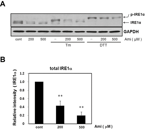
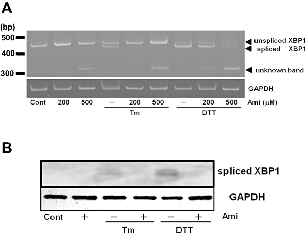
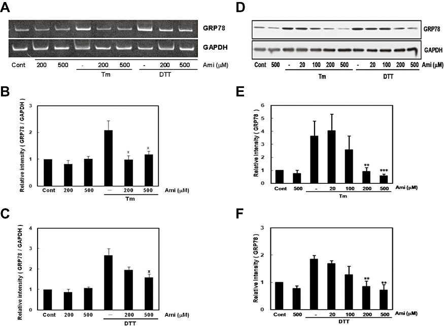
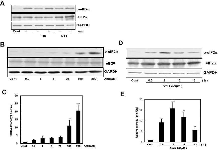
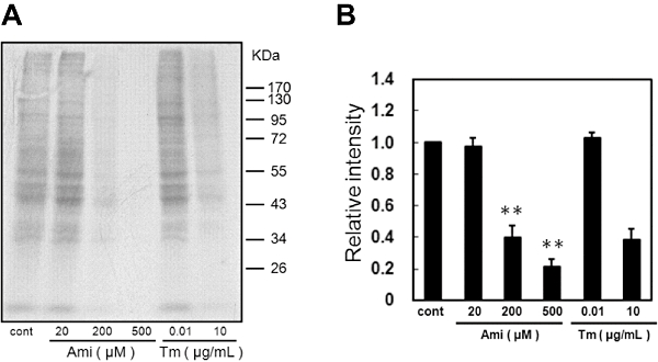
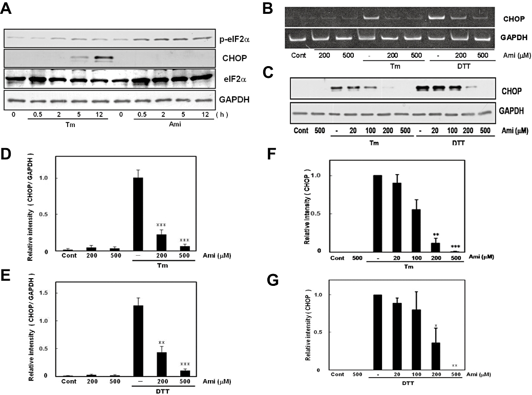
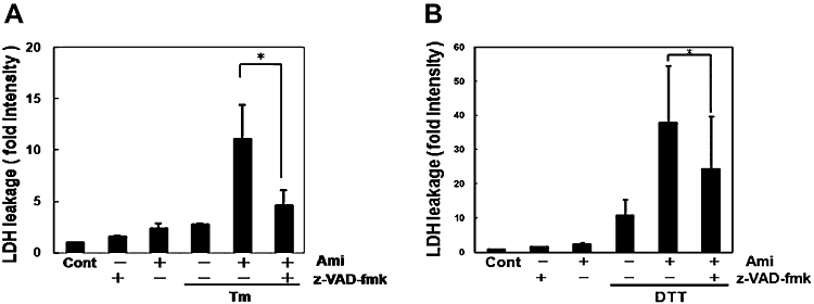
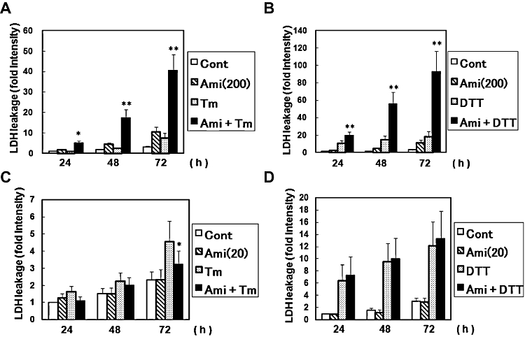
Key results: Amiloride completely inhibited ER stress-induced activation of IRE1alpha, an ER-localized stress sensor protein, splicing of XBP1, and subsequent expression of GRP78 at the mRNA and protein levels. ER stress induces the phosphorylation of eIF2alpha, leading to the expression of CHOP or an attenuation of translation in cells. Surprisingly, treatment with amiloride alone markedly promoted the phosphorylation but actually inhibited ER stress-induced CHOP expression. Finally, we found that amiloride (200 microM) synergistically enhanced ER stress-induced cell death, which was mediated through caspases. On the other hand, a low dose of amiloride (20 microM) significantly prevented Tm-induced cell death.

Conclusions and implications: These results suggest that amiloride can modulate UPR. They also suggest amiloride to be an important pharmacological agent and provide basic information for understanding and preventing ER stress-related diseases.


Figures

Figure 1
Figure 1
Effect of amiloride (Ami) on the expression of IRE1α and endoplasmic reticulum stress-induced IRE1α phosphorylation. (A) Cells were treated with Ami (200–500 µM) for 1 h and then stimulated with tunicamycin (Tm: 0.01 µg·mL−1) or dithiothreitol (DTT: 3 mM) for 5 h. Western blotting was performed using anti-IRE1α antibody. The band was detected at about 130 kDa∼ for normal and shifted band for IRE1α. Amiloride inhibited Tm-induced IRE1α phosphorylation. Amiloride inhibited the expression of IRE1α. (B) Densitometric analysis of total IRE1α expression using image analyzing software. **P < 0.01 versus control (n= 5). Cont, control.
Figure 2
Figure 2
Amiloride (Ami) inhibited endoplasmic reticulum stress-induced X-box binding protein 1 (XBP1) splicing. (A) Cells were treated with Ami (200–500 µM) for 1 h and then stimulated with tunicamycin (Tm: 0.01 µg·mL−1) or dithiothreitol (DTT: 3 mM) for 6 h. Reverse transcription-polymerase chain reaction was performed using specific primers for XBP1 and GAPDH. (B) Cells were treated with amiloride (Ami: 200 µM) for 1 h and then stimulated with tunicamycin (Tm: 0.01 µg·mL−1) or dithiothreitol (DTT: 3 mM) for 18 h. Western blotting was performed using anti-XBP1 (54 kDa) and anti-GAPDH (37 kDa) antibodies. Cont, control.
Figure 3
Figure 3
Amiloride (Ami) inhibited endoplasmic reticulum (ER) stress-induced glucose regulated protein 78 (GRP78) expression. (A) Cells were treated with Ami (20–500 µM) for 1 h and then stimulated with tunicamycin (Tm: 0.01 µg·mL−1) or dithiothreitol (DTT: 3 mM) for 6 h. Reverse transcription-polymerase chain reaction analysis was performed using specific primers for GRP78 and GAPDH. (B) and (C) Densitometric analysis of GRP78 mRNA using image analysing software. Ami dose-dependently inhibited ER stress-induced GRP78 expression. *P < 0.05, versus Tm or DTT (n= 5). (D) Cells were treated with Ami (20–500 µM) for 1 h and then stimulated with Tm (0.01 µg·mL−1) or DTT (3 mM) for 18 h. Western blotting was performed using anti-KDEL (78 kDa) or anti-GAPDH (37 kDa) antibodies. (E) and (F) Densitometric analysis of GRP78 protein using image analysing software. **P < 0.01, ***P < 0.001 versus Tm or DTT (n= 5). Cont, control.
Figure 4
Figure 4
Amiloride (Ami) increased eIF2α phosphorylation. (A) Mouse primary cultured glial cells were treated with Ami (200 µM) for 1 h and then stimulated with tunicamycin (Tm: 0.01 µg·mL−1) or dithiothreitol (DTT: 3 mM) for 5 h. (B) Cells were treated with Ami (0.2–200 µM) for 5 h. (D) Cells were treated with Ami (200 µM) for the periods indicated. Western blotting was performed using anti-phospho-eIF2α (42 kDa) and anti-GAPDH (37 kDa) antibodies. (C) and (E) Densitometric analysis of the phosphorylation of eIF2α using image analysing software. **P < 0.01, ***P < 0.001 versus control (n= 4 and 5 respectively). Cont, control.
Figure 5
Figure 5
Amiloride (Ami) inhibited translation. (A) Rates of protein synthesis were measured based on the incorporation of [35S]-methionine/cystine into proteins during a 30 min pulse of labelling followed by a 90 min exposure to the indicated concentrations of Ami. (B) Densitometric analysis of translation rates using image analysing software. **P < 0.01 versus control (n= 3). Tm, tunicamycin. Cont, control.
Figure 6
Figure 6
Amiloride (Ami) did not induce CCAAT/enhancer-binding protein homologous protein (CHOP) expression. (A) Cells were treated with tunicamycin (Tm: 0.01 µg·mL−1) or Ami (200 µM) for the periods indicated. Although Ami increased eIF2α phosphorylation, it did not induce CHOP expression. Western blotting was performed using anti-phospho-eIF2α (42 kDa), anti-eIF2α (40 kDa), anti-CHOP (29 kDa) and anti-GAPDH (37 kDa) antibodies. (B) Cells were treated with Ami (20–500 µM) for 1 h and then stimulated with Tm (0.01 µg·mL−1) or dithiothreitol (DTT: 3 mM) for 6 h. Reverse transcription-polymerase chain reaction was performed using specific primers for CHOP and GAPDH. (C) Cells were treated with Ami (20–500 µM) for 1 h and then stimulated with Tm (0.01 µg·mL−1) or DTT (3 mM) for 18 h. Western blotting was performed using anti-CHOP (29 kDa) and anti-GAPDH (37 kDa) antibodies. (D–G) Densitometric analysis of CHOP mRNA and protein using image analysing software. Ami dose-dependently inhibited ER stress-induced CHOP expression. *P < 0.05, **P < 0.01, ***P < 0.001 versus Tm or DTT (n= 6 for RT-PCR, n= 5 for Western blotting). Cont, control.
Figure 8
Figure 8
A caspase inhibitor prevented cell death caused by the combination of amiloride (Ami) and endoplasmic reticulum (ER) stress. (A) and (B) Cells were pretreated with the caspase inhibitor z-VAD-fmk (100 µM) for 30 min and treated with Ami (200 µM) for 1 h, and then stimulated with (A) tunicamycin (Tm: 0.1 µg·mL−1) or (B) dithiothreitol (DTT: 3 mM) for 72 h. Lactate dehydrogenase (LDH) activity was measured as an indicator of cytotoxicity. *P < 0.05 compared with Ami + ER stress (n= 3). Cont, control.
Figure 7
Figure 7
Effect of amiloride (Ami) on endoplasmic reticulum stress-induced cell death. (A) and (B) Cells were treated with Ami (200 µM) for 1 h and then stimulated with tunicamycin (Tm: 0.1 µg·mL−1) or dithiothreitol (DTT: 3 mM) for the periods indicated. Lactate dehydrogenase (LDH) activity was measured as an indicator of cytotoxicity. (C) and (D) Cells were treated with Ami (20 µM) for 1 h and then stimulated with Tm (0.1 µg mL−1) or DTT (3 mM) for the indicated times. *P < 0.05, **P < 0.01 Tm or DTT versus Ami + Tm or Ami + DTT (n= 3–8). Cont, control.

Similar articles

Cited by

References

    1. Aickin CC, Thomas RC. An investigation of the ionic mechanism of intracellular pH regulation in mouse soleus muscle fibres. J Physiol. 1977;273:295–316. - PMC - PubMed
    1. Alexander SPH, Mathie A, Peters JA. Guide to Receptors and Channels (GRAC), 3rd edition (2008 revision) Br J Pharmacol. 2008;153(Suppl. 2):S1–S209. - PMC - PubMed
    1. Arias RL, Sung ML, Vasylyev D, Zhang MY, Albinson K, Kubek K, et al. Amiloride is neuroprotective in an MPTP model of Parkinson's disease. Neurobiol Dis. 2008;31:334–341. - PubMed
    1. Bertolotti A, Zhang Y, Hendershot LM, Harding HP, Ron D. Dynamic interaction of BiP and ER stress transducers in the unfolded-protein response. Nat Cell Biol. 2000;2:326–332. - PubMed
    1. Boyce M, Bryant KF, Jousse C, Long K, Harding HP, Scheuner D, et al. A selective inhibitor of eIF2α dephosphorylation protects cells from ER stress. Science. 2005;307:935–939. - PubMed

Publication types

MeSH terms