Comparative Study
. 2010 May;160(2):185-98.
doi: 10.1111/j.1365-2249.2009.04074.x. Epub 2009 Dec 15.

An alpha-galactosylceramide C20:2 N-acyl variant enhances anti-inflammatory and regulatory T cell-independent responses that prevent type 1 diabetes

Affiliations
Comparative Study

An alpha-galactosylceramide C20:2 N-acyl variant enhances anti-inflammatory and regulatory T cell-independent responses that prevent type 1 diabetes

D Ly et al. Clin Exp Immunol. 2010 May.

Abstract

Protection from type 1 diabetes (T1D), a T helper type 1 (Th1)-mediated disease, is achievable in non-obese diabetic (NOD) mice by treatment with alpha-galactosylceramide (alpha-GalCer) glycolipids that stimulate CD1d-restricted invariant natural killer T (iNK T) cells. While we have reported previously that the C20:2 N-acyl variant of alpha-GalCer elicits a Th2-biased cytokine response and protects NOD mice from T1D more effectively than a form of alpha-GalCer that induces mixed Th1 and Th2 responses, it remained to determine whether this protection is accompanied by heightened anti-inflammatory responses. We show that treatment of NOD mice with C20:2 diminished the activation of 'inflammatory' interleukin (IL)-12 producing CD11c(high)CD8+ myeloid dendritic cells (mDCs) and augmented the function of 'tolerogenic' DCs more effectively than treatment with the prototypical iNKT cell activator KRN7000 (alpha-GalCer C26:0) that induces Th1- and Th2-type responses. These findings correlate with a reduced capacity of C20:2 to sustain the early transactivation of T, B and NK cells. They may also explain our observation that C20:2 activated iNK T cells depend less than KRN7000 activated iNK T cells upon regulation by regulatory T cells for cytokine secretion and protection from T1D. The enhanced anti-inflammatory properties of C20:2 relative to KRN7000 suggest that C20:2 should be evaluated further as a drug to induce iNK T cell-mediated protection from T1D in humans.


Figures
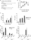
Fig. 1
Fig. 1
C20:2-activated invariant natural killer T (iNK T) cells induce a T helper type 2 (Th2)-biased cytokine response. (a) Structures of synthetic glycolipids α-galactosyl-ceramide (α-GalCer) C26:0 (KRN7000) and α-GalCer C20:2. Splenocytes from non-obese diabetic (NOD) mice (5–6 weeks old) were stimulated in vitro with the indicated concentrations of glycolipids for 72 h, and were then assayed by [3H]-thymidine incorporation for their proliferative capacity (b) or enzyme-linked immunosorbent assay (ELISA) for their cytokine secretion (c). Data show the means ± standard deviation (s.d.) for six replicates pooled from two independent experiments containing three mice per group. (d) NOD mice (5–6 weeks old) were injected once intravenously with 500 µg of anti-CD25 monoclonal antibody or immunoglobulin G (IgG), rested for 3 days, and then treated intraperitoneally with 4 µg of KRN7000, C20:2 or vehicle. Serum was collected from the mice at the indicated times post-treatment and analysed by ELISA for the concentrations of interleukin-4 and interferon-γ. Data show the means ± s.d. of three to five individual mice per group. *P < 0·01 compared to IgG/glycolipid treatment.
Fig. 2
Fig. 2
C20:2-activated invariant natural killer T (iNK T) cells depend less on CD4+CD25+ regulatory T cell (Treg) activity than KRN7000-activated iNK T cells for protection against type 1 diabetes (T1D). Non-obese diabetic (NOD) mice (4–5 weeks old) were administered anti-CD25 or immunoglobulin G (IgG), rested, and then treated with a multi-low-dose protocol of KRN7000, C20:2 or vehicle (4 µg/dose, every other day for 3 weeks). The incidence of T1D in these mice was monitored to 30 weeks of age. Results show the cumulative incidence of T1D from two independent experiments conducted using four to six mice per group. *P < 0·04 compared to vehicle-treated mice.
Fig. 3
Fig. 3
Kinetics of invariant natural killer T (iNK T) cell expansion activated with KRN7000 or C20:2. Non-obese diabetic (NOD) mice (4–5 weeks old) were administered anti-CD25 or immunoglobulin G (IgG), rested, and then treated with KRN7000, C20:2 or vehicle, as in Fig. 1. Spleen (a) or pancreatic lymph nodes (PLN) (b) lymphocytes were harvested at the indicated time-points post-glycolipid treatment for flow cytometric analysis of their frequency of iNK T cells. Dot plots from a representative experiment are shown with the means ± standard deviation (s.d.) of gated [T cell receptor (TCR)-β+ and α-galactosyl-ceramide C26:0 (α-GalCer)-CD1d tetramer+] iNK T cells from three or five individual mice per group (a and b, left panel). Absolute numbers of iNK T cells observed at 3 days post-glycolipid treatment are shown for each group (a and b, right panel). Data represent the means ± s.d. from three individual mice per group. *P < 0·001 compared to IgG for a given glycolipid treatment.
Fig. 4
Fig. 4
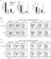
C20:2 restimulation induces T helper type 2 (Th2) cytokine production by invariant natural killer T (iNK T) cells but does not polarize iNK T cells to a Th2-type response. Non-obese diabetic (NOD) mice (4–5 weeks old) were administered anti-CD25 or immunoglobulin G (IgG), rested, and then treated with KRN7000, C20:2 or vehicle, as in Fig. 1. (a) Splenocytes harvested at 3 days post-glycolipid treatment were restimulated in vitro for 72 h with KRN7000 or C20:2 (100 ng/ml), or vehicle, and their levels of cytokine [interferon (IFN)-γ, interleukin (IL)-4, IL-13] secretion were assayed. Data represent the means ± SD of triplicate measurements. Representative data from two independent experiments containing three mice per group are shown. (b) Spleen and PLN lymphocytes harvested from mice at 2 h post-glycolipid treatment were cultured in vitro for 3 h in the presence of a protein transport inhibitor Golgi Stop, and stained for intracellular IL-4 and IFN-γ in gated CD4+ iNK T cells. Representative pseudocolour contour plots from three independent and reproducible experiments are shown. Percentages of total cells in each quadrant are indicated.
Fig. 5
Fig. 5
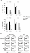
C20:2-activated invariant natural killer T (iNK T) cells show a reduced ability to sustain the transactivation of B cells and NK cells. Non-obese diabetic (NOD) mice (4–5 weeks old) were administered anti-CD25 or immunoglobulin G (IgG), rested, and then treated with KRN7000, C20:2 or vehicle, as in Fig. 1. (a) Splenocytes were harvested at the indicated time-points post-glycolipid treatment, gated on B cells (B220+CD3) or NK cells (DX5+CD3-) and the frequency of CD69+ cells was analysed by flow cytometry. Data represent the means ± standard deviation (s.d.) of three or four individual mice per group. (b) Splenocytes harvested from the mice at the indicated time-points post-glycolipid treatment were cultured in vitro for 3 h in the presence of a protein transport inhibitor Golgi Stop. Gated NK cells (DX5+CD3) were stained for intracellular interferon (IFN)-γ and representative contour plots from two or three independent and reproducible experiments are shown. The percentage of IFN-γ+ cells in the gated NK cell populations are indicated. *P < 0·01 compared between glycolipids for a given anti-CD25 or IgG treatment; **P < 0·05 compared between glycolipids for a given treatment group.
Fig. 6
Fig. 6
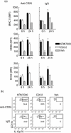
Both C20:2- and KRN7000-activated invariant natural killer T (iNK T) cells induce dendritic cell (DC) maturation but only KRN7000 sustains this maturation. Non-obese diabetic (NOD) mice (4–5 weeks old) were administered anti-CD25 or immunoglobulin G (IgG), rested, and then treated with KRN7000, C20:2 or vehicle, as in Fig. 1. Splenocytes were harvested at the indicated time-points post-glycolipid treatment. (a) CD11chigh mDC were gated and the mean fluorescence intensity (MFI) of the complete FL-2 channel staining for major histocompatibility complex (MHC) class II, CD86 and CD40 were graphed for the 6 h and 24 h time-points after glycolipid treatment. Data represent the means ± standard deviation (s.d.) of four individual mice per group. (b) At 6 h post-treatment with glycolipid, splenocytes were harvested, cultured in vitro for 3 h in the presence of a protein transport inhibitor, Golgi Stop, and CD11chighCD8+ mDCs (Siglec HCD11chigh) were then gated and stained for intracellular interleukin-12. Representative contour plots from one of two independent and reproducible experiments are shown. *P < 0·01 compared between glycolipids for a given treatment group; **P < 0·05 compared between glycolipids for a given treatment group.
Fig. 7
Fig. 7
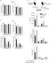
Activation of invariant natural killer T (iNK T) cells with C20:2 alters the frequency and function of dendritic cells (DCs). Non-obese diabetic (NOD) mice (4–5 weeks old) were administered anti-CD25 or immunoglobulin G (IgG), rested, and then treated with KRN7000, C20:2 or vehicle, as in Fig. 2. Splenocytes and pancreatic lymph nodes (PLN) were collected at 1 week after the last glycolipid dose. (a) Plasmacytoid DCs (pDCs) (Siglec H+CD11clow) and myeloid DCs (mDCs) (Siglec HCD11chigh) were gated as indicated for DC subset analysis, and mDCs were separated further into the CD11chighCD8 and CD11chighCD8+ subsets. (b) Splenocytes and PLN were gated as in (a), and the frequencies and PLN absolute number of the various DC subsets were analysed. Data shown represent the means ± standard deviation (s.d.) of five mice per group. (c) NOD mice (4–5 weeks old) were treated with a multi-low-dose protocol of KRN7000 or C20:2 (4 µg/dose, every other day for 3 weeks), or vehicle. One week after the last dose, spleen and PLN suspensions were pooled and CD11c+ DCs from each group were sorted. Concurrently, the Ins B9-23 peptide emulsified in incomplete Freund's adjuvant (IFA) or vehicle was administered to a separate group of NOD mice to prime their CD4+ T cells. Fluorescence activated cell sorted (FACS) Ins B9-23 peptide primed-CD4+ T cells were cultured with CD11c+ DCs in the presence of Ins B9-23 for 72 h. T cell proliferation and enzyme-linked immunosorbent assay (ELISA) assays of cytokine concentrations [interleukin (IL)-2, interferon (IFN)-γ, IL-4] in culture supernatants were then performed. *P < 0·05 compared to vehicle within a given treatment group; **P < 0·05 compared to C20:2 within a given treatment group.

Similar articles

Cited by

References

    1. Bendelac A, Savage PB, Teyton L. The biology of NKT cells. Annu Rev Immunol. 2007;25:297–336. - PubMed
    1. Kronenberg M. Toward an understanding of NKT cell biology: progress and paradoxes. Annu Rev Immunol. 2005;23:877–900. - PubMed
    1. Van Kaer L. Alpha-galactosylceramide therapy for autoimmune diseases: prospects and obstacles. Nat Rev Immunol. 2005;5:31–42. - PubMed
    1. Godfrey DI, MacDonald HR, Kronenberg M, Smyth MJ, Van Kaer L. NKT cells: what's in a name? Nat Rev Immunol. 2004;4:231–7. - PubMed
    1. Anderson MS, Bluestone JA. The NOD mouse: a model of immune dysregulation. Annu Rev Immunol. 2005;23:447–85. - PubMed

Publication types

MeSH terms