

Advances in imaging of new targets for pharmacological intervention in stroke: real-time tracking of T-cells in the ischaemic brain
- PMID: 20015295
- PMCID: PMC2829206
- DOI: 10.1111/j.1476-5381.2009.00527.x
Advances in imaging of new targets for pharmacological intervention in stroke: real-time tracking of T-cells in the ischaemic brain
Abstract
Background and purpose: T-cells may play a role in the evolution of ischaemic damage and repair, but the ability to image these cells in the living brain after a stroke has been limited. We aim to extend the technique of real-time in situ brain imaging of T-cells, previously shown in models of immunological diseases, to models of experimental stroke.
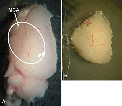
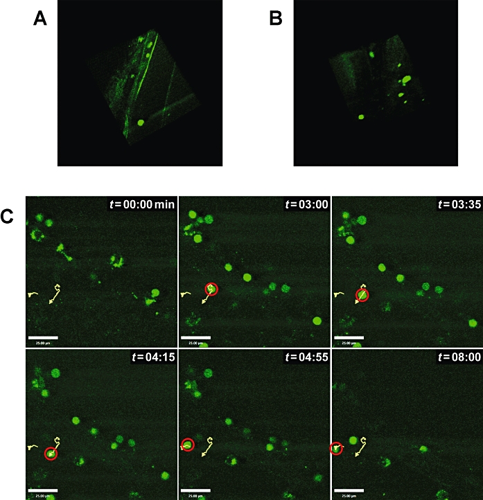
Experimental approach: Male C57BL6 mice (6-8 weeks) (n= 3) received a total of 2-5 x 10(6) carboxyfluorescein diacetate succinimidyl ester (CFSE)-labelled lymphocytes from donor C57BL6 mice via i.v. injection by adoptive transfer. Twenty-four hours later, recipient mice underwent permanent left distal middle cerebral artery occlusion (MCAO) by electrocoagulation or by sham surgery under isoflurane anaesthesia. Female hCD2-green fluorescent protein (GFP) transgenic mice that exhibit GFP-labelled T-cells underwent MCAO. At 24 or 48 h post-MCAO, a sagittal brain slice (1500 microm thick) containing cortical branches of the occluded middle cerebral artery (MCA) was dissected and used for multiphoton laser scanning microscopy (MPLSM).
Key results: Our results provide direct observations for the first time of dynamic T-cell behaviour in living brain tissue in real time and herein proved the feasibility of MPLSM for ex vivo live imaging of immune response after experimental stroke.
Conclusions and implications: It is hoped that these advances in the imaging of immune cells will provide information that can be harnessed to a therapeutic advantage.
Figures
Similar articles
-
In vivo real-time multiphoton imaging of T lymphocytes in the mouse brain after experimental stroke.Stroke. 2011 May;42(5):1429-36. doi: 10.1161/STROKEAHA.110.603704. Epub 2011 Mar 24. Stroke. 2011. PMID: 21441145
-
Visualizing chemokine-dependent T cell activation and migration in response to central nervous system infection.Methods Mol Biol. 2013;1013:171-83. doi: 10.1007/978-1-62703-426-5_11. Methods Mol Biol. 2013. PMID: 23625499 Free PMC article.
-
Tracking in vivo migration and distribution of antigen-specific cytotoxic T lymphocytes by 5,6-carboxyfluorescein diacetate succinimidyl ester staining during cancer immunotherapy.Chin Med J (Engl). 2013 Aug;126(16):3019-25. Chin Med J (Engl). 2013. PMID: 23981604
-
Carboxyfluorescein diacetate succinimidyl ester and the virgin lymphocyte: a marriage made in heaven.Immunol Cell Biol. 1999 Dec;77(6):530-8. doi: 10.1046/j.1440-1711.1999.00871.x. Immunol Cell Biol. 1999. PMID: 10571674 Review.
-
Divided we stand: tracking cell proliferation with carboxyfluorescein diacetate succinimidyl ester.Immunol Cell Biol. 1999 Dec;77(6):509-15. doi: 10.1046/j.1440-1711.1999.00864.x. Immunol Cell Biol. 1999. PMID: 10571671 Review.
Cited by
-
Themed section: Imaging in pharmacology.Br J Pharmacol. 2010 Feb;159(4):735-7. doi: 10.1111/j.1476-5381.2010.00685.x. Br J Pharmacol. 2010. PMID: 20388127 Free PMC article.
-
Neuroimmunological blood brain barrier opening in experimental cerebral malaria.PLoS Pathog. 2012;8(10):e1002982. doi: 10.1371/journal.ppat.1002982. Epub 2012 Oct 25. PLoS Pathog. 2012. PMID: 23133375 Free PMC article.
-
Adaptive Immunity Regulation and Cerebral Ischemia.Front Immunol. 2020 May 12;11:689. doi: 10.3389/fimmu.2020.00689. eCollection 2020. Front Immunol. 2020. PMID: 32477327 Free PMC article. Review.
-
Imaging--the interface with pharmacology: looking to the future.Br J Pharmacol. 2011 Aug;163(8):1563-4. doi: 10.1111/j.1476-5381.2011.01294.x. Br J Pharmacol. 2011. PMID: 21790531 Free PMC article.
-
Temporal dynamics of peripheral neutrophil and lymphocytes following acute ischemic stroke.Neurol Sci. 2019 Sep;40(9):1877-1885. doi: 10.1007/s10072-019-03919-y. Epub 2019 May 8. Neurol Sci. 2019. PMID: 31069585 Free PMC article.
References
-
- de Boer J, Williams A, Skavdis G, Harker N, Coles M, Tolaini M, et al. Transgenic mice with hematopoietic and lymphoid specific expression of Cre. Eur J Immunol. 2003;33:314–325. - PubMed
-
- Carswell HV, Dominiczak AF, Garcia-Segura LM, Harada N, Hutchison JB, Macrae IM. Brain aromatase expression after experimental stroke: topography and time course. J Steroid Biochem Mol Biol. 2005;96:89–91. - PubMed
-
- Flügel A, Odoardi F, Nosov M, Kawakami N. Autoaggressive effector T cells in the course of experimental autoimmune encephalomyelitis visualized in the light of two-photon microscopy. J Neuroimmunol. 2007;191:86–97. - PubMed
Publication types
MeSH terms
Substances
Grants and funding
LinkOut - more resources
Full Text Sources
