

A weak DD-carboxypeptidase activity explains the inability of PBP 6 to substitute for PBP 5 in maintaining normal cell shape in Escherichia coli
- PMID: 20015336
- PMCID: PMC3013634
- DOI: 10.1111/j.1574-6968.2009.01863.x
A weak DD-carboxypeptidase activity explains the inability of PBP 6 to substitute for PBP 5 in maintaining normal cell shape in Escherichia coli
Abstract
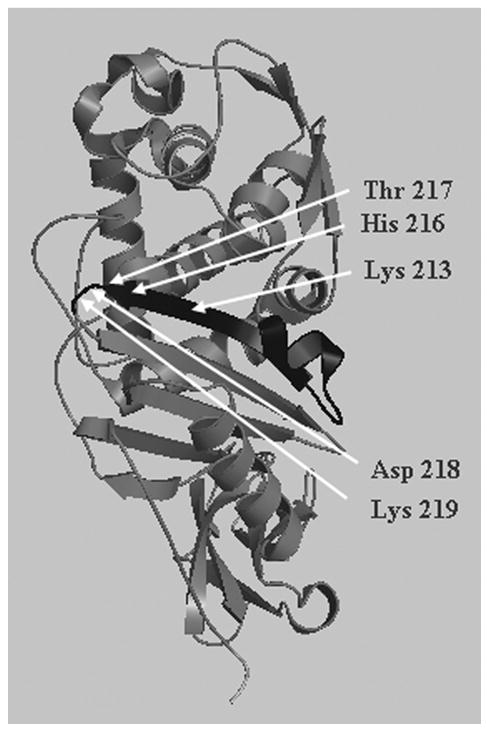
Penicillin-binding protein (PBP) 5 plays a critical role in maintaining normal cellular morphology in mutants of Escherichia coli lacking multiple PBPs. The most closely related homologue, PBP 6, is 65% identical to PBP 5, but is unable to substitute for PBP 5 in returning these mutants to their wild-type shape. The relevant differences between PBPs 5 and 6 are localized in a 20-amino acid stretch of domain I in these proteins, which includes the canonical KTG motif at the active site. We determined how these differences affected the enzymatic properties of PBPs 5 and 6 toward beta-lactam binding and the binding and hydrolysis of two peptide substrates. We also investigated the enzymatic properties of recombinant fusion proteins in which active site segments were swapped between PBPs 5 and 6. The results suggest that the in vivo physiological role of PBP 5 is distinguished from PBP 6 by the higher degree of DD-carboxypeptidase activity of the former.
Figures
References
-
- Amanuma H, Strominger JL. Purification and properties of penicillin-binding proteins 5 and 6 from Escherichia coli membranes. J Biol Chem. 1980;255:1173–1180. - PubMed
-
- Andrade MA, Chacon P, Merelo JJ, Moran F. Evaluation of secondary structure of proteins from UV circular dichroism spectra using an unsupervised learning neural network. Protein Eng. 1993;6:83–90. - PubMed
-
- Davies C, White SW, Nicholas RA. Crystal structure of a deacylation-defective mutant of penicillin-binding protein 5 at 2.3-A resolution. J Biol Chem. 2001;276:616–623. - PubMed
Publication types
MeSH terms
Substances
Grants and funding
LinkOut - more resources
Full Text Sources
Molecular Biology Databases
Research Materials
Miscellaneous
