. 2010 Feb 15;19(4):634-47.
doi: 10.1093/hmg/ddp531. Epub 2009 Dec 16.

Specific saposin C deficiency: CNS impairment and acid beta-glucosidase effects in the mouse

Affiliations

Specific saposin C deficiency: CNS impairment and acid beta-glucosidase effects in the mouse

Ying Sun et al. Hum Mol Genet. .

Abstract

Saposins A, B, C and D are derived from a common precursor, prosaposin (psap). The few patients with saposin C deficiency develop a Gaucher disease-like central nervous system (CNS) phenotype attributed to diminished glucosylceramide (GC) cleavage activity by acid beta-glucosidase (GCase). The in vivo effects of saposin C were examined by creating mice with selective absence of saposin C (C-/-) using a knock-in point mutation (cysteine-to-proline) in exon 11 of the psap gene. In C-/- mice, prosaposin and saposins A, B and D proteins were present at near wild-type levels, but the saposin C protein was absent. By 1 year, the C-/- mice exhibited weakness of the hind limbs and progressive ataxia. Decreased neuromotor activity and impaired hippocampal long-term potentiation were evident. Foamy storage cells were observed in dorsal root ganglion and there was progressive loss of cerebellar Purkinje cells and atrophy of cerebellar granule cells. Ultrastructural analyses revealed inclusions in axonal processes in the spinal cord, sciatic nerve and brain, but no excess of multivesicular bodies. Activated microglial cells and astrocytes were present in thalamus, brain stem, cerebellum and spinal cord, indicating regional pro-inflammatory responses. No storage cells were found in visceral organs of these mice. The absence of saposin C led to moderate increases in GC and lactosylceramide (LacCer) and their deacylated analogues. These results support the view that saposin C has multiple roles in glycosphingolipid (GSL) catabolism as well as a prominent function in CNS and axonal integrity independent of its role as an optimizer/stabilizer of GCase.


Figures

Figure 1.
Figure 1.
Targeting construct and verification. (A) Schematic map of the saposin C targeting construct. Individual saposins are encoded by the exons underlined with a dotted line. The mutation in the saposin C (Cys→Pro) domain was in prosaposin exon 11. The point mutation destroyed one of three disulfide bridges within saposin C, which leads to a deficiency in saposin C protein. The neo gene was removed by recombination of two loxP sites by cross-breeding of saposin C heterozygotes and Zp3-Cre transgenic mice. B, BamHI; Bgl, BglII; H, HindIII; E, EcoRI; Ev, EcoRV; X, XbaI; Xho, XhoI; Nhe, NheI; Sal, SalI; *mutation site. (B) Correct targeting of ES cells was confirmed by Southern blot. The clones were digested by HindIII for 5′ probe and EcoRI for 3′ probe. (C) PCR genotyping of saposin C mice before and after Cre recombination using primers C–F and C–R across the loxP site.
Figure 2.
Figure 2.
Saposin protein expression. (A) Saposin C was not detected in the fibroblasts using anti-mouse saposin C antibody. Saposin A and B were detected by anti-mouse saposin A and B antibodies, respectively. (B) Prosaposin and saposin D were detected with anti-mouse saposin D antibody. Prosaposin levels in saposin C−/− fibroblasts were slightly increased compared to the WT control. β-Actin was used as a loading control.
Figure 3.
Figure 3.
Phenotype of saposin C−/− mice at 16 months. (Left) Saposin C−/− mouse showing hind paw spreading suggesting paralysis. (Right) Saposin C−/− mouse with abnormal hindleg positioning and WT mouse with a normal V shape position in tail-hanging reflex.
Figure 4.
Figure 4.
Neurobehavioral assessment. (A) Narrow bridges test. WT and saposin C−/− mice were tested on 11, 17 and 28 mm round beams at 9 and 13 months. Saposin C−/− mice showed significant increases of latency (left) across the beam and foot slips (right) compared with WT. Latency and number of slips in saposin C−/− mice progressively increased with age. (B) Zero-maze test. Relative to WT, saposin C−/− mice had fewer entries and spent longer time in the open area. (C) Locomotor activity. (Left) Saposin C−/− mice exhibited significantly enhanced vertical activity (P < 0.02) compared with WT. (Right) Total distance activity in saposin C−/− mice was not different from WT (P < 0.1). Activity was recorded every 5 min over a 60-min period. WT and saposin C−/− male mice were used in the tests. *P ≤ 0.05; **P ≤ 0.01; ***P ≤ 0.001. WT, n = 16; saposin C−/−, n = 21.
Figure 5.
Figure 5.
Electrophysiology analysis of long-term potentiation (LTP). The fEPSPs were recorded from parasagittal sections (350 µm) of hippocampal CA1 region using the MED64 multielectrode array system to determine the slope of resulting EPSPs. The slopes of the EPSPs (LTP) in 24-month-old saposin C−/− mice (closed circle) were significantly (P < 0.05) decreased compared with WT mice (open circle) for 90 min following stimulation. WT, n = 7 mice; saposin C−/−, n = 6 mice.
Figure 6.
Figure 6.
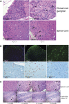
CNS pathology in C−/− mice. (A) The paraffin sections of dorsal root ganglion and dorsal horn of spinal cord from 15-month-old saposin C+/− or C−/− mice were stained with H&E. Foamy storage material (arrows) was present in the dorsal root ganglion neurons of saposin C−/− mice. Larger axonal spheroids (arrows) were present in the spinal cord dorsal horn of these mice. As a control, saposin C+/− mice had normal morphology in dorsal root ganglion and spinal cord. (B) CNS proinflammation in saposin C−/− mice demonstrated by anti-GFAP (B1-B3) and anti-CD68 (B4-B6). (B1) WT spinal cord, (B2) saposin C−/− spinal cord and (B3) C−/− midbrain at 15 months showed astrogliosis with enhanced GFAP (green) signal. The nucleus was labeled by DAPI (blue). (B4) C−/− dorsal horn of spinal cord. (B5) C−/− thalamus. (B6) C−/− brain stem. Microglial cells were stained by anti-CD68 antibody (brown) in 24-month-old saposin C−/− mice. Tissue sections were counterstained with hematoxylin. (C) H&E stained paraffin sections of spinal cord and cerebellum from saposin C−/− mice at 3, 6, 12 and 18 months. Axonal spheroids were present in dorsal horn of spinal cord at 6, 12 and 18 months, but absent at 3 months. Purkinje cell layer (arrows) in cerebella lobule IV was normal at 3 months. Loss of Purkinje cells (*) was evident at 6 months and older.
Figure 7.
Figure 7.
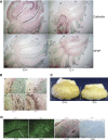
Pathology in cerebellum. (A) Anti-calbindin antibody stained Purkinje cells (brown) in 24-month-old C+/− and C−/− mouse brain. Loss of Purkinje cells in saposin C−/− mice progressed from cerebella lobule III to X with age. Increases of astrogliosis demonstrated by anti-GFAP antibody staining (brown) were in the region where the Purkinje cells were lost. Normal Purkinje cell layers and astrocytes were found in the C+/− cerebellum. (B) Calbindin staining revealed axon and neuronal processes in the molecular layer and white matter of the cerebellum. (B1) Normal Purkinje cells and axons (arrows) in C+/− mouse. (B2) Degeneration of Purkinje cell axons (stars) in the molecular layer of cerebella lobule X in 24-month-old C−/− mouse. (B3) Neuronal processes in C+/− cerebella white matter. (B4) Neural spheroids (arrows) presented in the neuronal processes of C−/− mouse. (C) 24-month-old C+/− and C−/− mouse cerebellum. The white bars show the height of cerebellum. The size of saposin C−/− cerebellum was reduced at 24 months. (D) Atrophy in the granule cell layer of saposin C−/− mice. NeuN staining cells (green) were decreased in the granulecell layer of cerebella lobule III from 24-month-old C−/− mice compared with lobule III of C+/− mouse. The white bar in the photo shows the diameter of the granule cell layer. Both GFAP (brown) and CD68 (brown) signals were enhanced in lobule III of the saposin C−/− mouse. Sections stained with calbindin and GFAP were counterstained with methyl green (green). CD68-stained sections were counterstained with hemotoxilin.
Figure 8.
Figure 8.
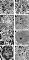
Ultrastructural studies. (A) The spinal cord of saposin C+/− mice at 18 months had normal morphology. (B) The spinal cord of saposin C−/− mice at 18 months showed inclusion bodies (arrows) in neuronal process. (C) Enlarged view of inclusion bodies in (B). (D) Cortical neuron in saposin C−/− mice at 10 months had normal morphology. (E) Axonal inclusions (arrow) in granule cell layer of saposin C−/− cerebellum at 18 months. (F) Purkinje cells in saposin C−/− mouse at 10 months had normal morphology. (G) The sciatic nerve of saposin C−/− mice at 18 months had inclusion materials (arrows) and degenerating myelin layers (arrowheads). (H) Axonal inclusion material (arrow) in the midbrain of saposin C−/− mice at 18 months.
Figure 9.
Figure 9.
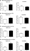
GSL analyses. Spinal cord and cerebellum lysate were analyzed for GC, GS, LacCer and LacSph. Saposin C−/− mice had significant increases of GC, GS and LacSph in the spinal cord. LacCer and LacSph increased in the C−/− cerebellum. The amounts of lipids were normalized to phosphate in each sample. Age-matched 13-month-old WT and C−/− mice were used in the assay (n = 3). *P ≤ 0.05 and **P ≤ 0.01.
Figure 10.
Figure 10.
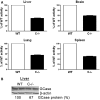
GCase activity and protein in C−/− mice. (A) GCase activity levels were decreased in C−/− liver, lung, brain and spleen by ∼50% relative to the WT level. (B) The GCase protein level in the C−/− liver was 67% of the WT level. Age-matched 2-month-old WT and C−/− mice were used in the assay (n = 3).

Similar articles

Cited by

References

    1. Leonova T., Qi X., Bencosme A., Ponce E., Sun Y., Grabowski G.A. Proteolytic processing patterns of prosaposin in insect and mammalian cells. J. Biol. Chem. 1996;271:17312–17320. - PubMed
    1. Sandhoff K., Kolter T., Van Echten-Deckert G. Sphingolipid metabolism. Sphingoid analogs, sphingolipid activator proteins, and the pathology of the cell. Ann. NY Acad. Sci. 1998;845:139–151. - PubMed
    1. Li S.C., Sonnino S., Tettamanti G., Li Y.T. Characterization of a nonspecific activator protein for the enzymatic hydrolysis of glycolipids. J. Biol. Chem. 1988;263:6588–6591. - PubMed
    1. Sun Y., Witte D.P., Ran H., Zamzow M., Barnes S., Cheng H., Han X., Williams M.T., Skelton M.R., Vorhees C.V., et al. Neurological deficits and glycosphingolipid accumulation in saposin B deficient mice. Hum. Mol. Genet. 2008;17:2345–2356. - PMC - PubMed
    1. Zhang X.L., Rafi M.A., DeGala G., Wenger D.A. Insertion in the mRNA of a metachromatic leukodystrophy patient with sphingolipid activator protein-1 deficiency. Proc. Natl Acad. Sci. USA. 1990;87:1426–1430. - PMC - PubMed

Publication types

MeSH terms