Extracellular ubiquitin inhibits beta-AR-stimulated apoptosis in cardiac myocytes: role of GSK-3beta and mitochondrial pathways
- PMID: 20015977
- PMCID: PMC2836259
- DOI: 10.1093/cvr/cvp402
Extracellular ubiquitin inhibits beta-AR-stimulated apoptosis in cardiac myocytes: role of GSK-3beta and mitochondrial pathways
Abstract
Aims: Beta-adrenergic receptor (beta-AR) stimulation induces apoptosis in adult rat ventricular myocytes (ARVMs) via the activation of glycogen synthase kinase-3beta (GSK-3beta) and mitochondrial pathways. However, beta-AR stimulation induces apoptosis only in a fraction ( approximately 15-20%) of ARVMs. We hypothesized that ARVMs may secrete/release a survival factor(s) which protects 80-85% of cells from apoptosis.
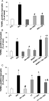
Methods and results: Using two-dimensional gel electrophoresis followed by MALDI TOF and MS/MS, we identified ubiquitin (Ub) in the conditioned media of ARVMs treated with beta-AR agonist (isoproterenol). Western blot analysis confirmed increased Ub levels in the conditioned media 3 and 6 h after beta-AR stimulation. Inhibition of beta1-AR and beta2-AR subtypes inhibited beta-AR-stimulated increases in extracellular levels of Ub, whereas activation of adenylyl cyclase using forskolin mimicked the effects of beta-AR stimulation. Incubation of cells with exogenous biotinylated Ub followed by western blot analysis of the cell lysates showed uptake of extracellular Ub into cells, which was found to be higher after beta-AR stimulation (1.9 +/- 0.4-fold; P < 0.05 vs. control, n = 6). Pre-treatment with Ub inhibited beta-AR-stimulated increases in apoptosis. Inhibition of phosphoinositide 3-kinase using wortmannin and LY-294002 prevented anti-apoptotic effects of extracellular Ub. Ub pre-treatment inhibited beta-AR-stimulated activation of GSK-3beta and c-Jun N-terminal kinase (JNK) and increases in the levels of cytosolic cytochrome c. The use of methylated Ub suggested that the anti-apoptotic effects of extracellular Ub are mediated via monoubiquitination.
Conclusion: beta-AR stimulation increases levels of Ub in the conditioned media. Extracellular Ub plays a protective role in beta-AR-stimulated apoptosis, possibly via the inactivation of GSK-3beta/JNK and mitochondrial pathways.
Figures


Comment in
-
Ubiquitin, a novel paracrine messenger of cardiac cell survival.Cardiovasc Res. 2010 Apr 1;86(1):1-3. doi: 10.1093/cvr/cvq026. Epub 2010 Jan 25. Cardiovasc Res. 2010. PMID: 20100704 Free PMC article. No abstract available.
Similar articles
-
NF2 signaling pathway plays a pro-apoptotic role in β-adrenergic receptor stimulated cardiac myocyte apoptosis.PLoS One. 2018 Apr 30;13(4):e0196626. doi: 10.1371/journal.pone.0196626. eCollection 2018. PLoS One. 2018. PMID: 29709009 Free PMC article.
-
Glycogen synthase kinase-3beta plays a pro-apoptotic role in beta-adrenergic receptor-stimulated apoptosis in adult rat ventricular myocytes: Role of beta1 integrins.J Mol Cell Cardiol. 2007 Mar;42(3):653-61. doi: 10.1016/j.yjmcc.2006.12.011. Epub 2006 Dec 29. J Mol Cell Cardiol. 2007. PMID: 17292911 Free PMC article.
-
beta-Adrenergic receptor-stimulated apoptosis in adult cardiac myocytes involves MMP-2-mediated disruption of beta1 integrin signaling and mitochondrial pathway.Am J Physiol Cell Physiol. 2006 Jan;290(1):C254-61. doi: 10.1152/ajpcell.00235.2005. Epub 2005 Sep 7. Am J Physiol Cell Physiol. 2006. PMID: 16148033
-
Extracellular Ubiquitin: Role in Myocyte Apoptosis and Myocardial Remodeling.Compr Physiol. 2015 Dec 15;6(1):527-60. doi: 10.1002/cphy.c150025. Compr Physiol. 2015. PMID: 26756642 Review.
-
The control of cardiomyocyte apoptosis via the beta-adrenergic signaling pathways.Arch Mal Coeur Vaiss. 2005 Mar;98(3):236-41. Arch Mal Coeur Vaiss. 2005. PMID: 15816327 Review.
Cited by
-
Exogenous ubiquitin modulates chronic β-adrenergic receptor-stimulated myocardial remodeling: role in Akt activity and matrix metalloproteinase expression.Am J Physiol Heart Circ Physiol. 2012 Dec 15;303(12):H1459-68. doi: 10.1152/ajpheart.00401.2012. Epub 2012 Oct 5. Am J Physiol Heart Circ Physiol. 2012. PMID: 23042947 Free PMC article.
-
Cardioprotective Potential of Exogenous Ubiquitin.Cardiovasc Drugs Ther. 2021 Dec;35(6):1227-1232. doi: 10.1007/s10557-020-07042-5. Epub 2020 Sep 10. Cardiovasc Drugs Ther. 2021. PMID: 32910339 Free PMC article. Review.
-
NF2 signaling pathway plays a pro-apoptotic role in β-adrenergic receptor stimulated cardiac myocyte apoptosis.PLoS One. 2018 Apr 30;13(4):e0196626. doi: 10.1371/journal.pone.0196626. eCollection 2018. PLoS One. 2018. PMID: 29709009 Free PMC article.
-
Ubiquitin is a versatile scaffold protein for the generation of molecules with de novo binding and advantageous drug-like properties.FEBS Open Bio. 2015 Jul 10;5:579-93. doi: 10.1016/j.fob.2015.07.002. eCollection 2015. FEBS Open Bio. 2015. PMID: 26258013 Free PMC article.
-
Increased extracellular ubiquitin in surgical wound fluid provides a chemotactic signal for myeloid dendritic cells.Eur J Trauma Emerg Surg. 2020 Feb;46(1):153-163. doi: 10.1007/s00068-018-1001-0. Epub 2018 Aug 30. Eur J Trauma Emerg Surg. 2020. PMID: 30159662
References
-
- Andreka P, Nadhazi Z, Muzes G, Szantho G, Vandor L, Konya L, et al. Possible therapeutic targets in cardiac myocyte apoptosis. Curr Pharm Des. 2004;10:2445–2461. - PubMed
-
- Singh K, Xiao L, Remondino A, Sawyer DB, Colucci WS. Adrenergic regulation of cardiac myocyte apoptosis. J Cell Physiol. 2001;189:257–265. - PubMed
-
- Iwai-Kanai E, Hasegawa K, Araki M, Kakita T, Morimoto T, Sasayama S. Alpha- and beta-adrenergic pathways differentially regulate cell type-specific apoptosis in rat cardiac myocytes. Circulation. 1999;100:305–311. - PubMed
-
- Zaugg M, Xu W, Lucchinetti E, Shafiq SA, Jamali NZ, Siddiqui MA. Beta-adrenergic receptor subtypes differentially affect apoptosis in adult rat ventricular myocytes. Circulation. 2000;102:344–350. - PubMed
-
- Shizukuda Y, Buttrick PM, Geenen DL, Borczuk AC, Kitsis RN, Sonnenblick EH. Beta-adrenergic stimulation causes cardiocyte apoptosis: influence of tachycardia and hypertrophy. Am J Physiol. 1998;275:H961–H968. - PubMed
Publication types
MeSH terms
Substances
Grants and funding
LinkOut - more resources
Full Text Sources
Research Materials
Miscellaneous

