Role of untranslated regions in regulation of gene expression, replication, and pathogenicity of Newcastle disease virus expressing green fluorescent protein
- PMID: 20015997
- PMCID: PMC2820931
- DOI: 10.1128/JVI.02049-09
Role of untranslated regions in regulation of gene expression, replication, and pathogenicity of Newcastle disease virus expressing green fluorescent protein
Abstract
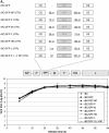
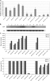
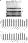
To gain insight into the role of untranslated regions (UTRs) in regulation of foreign gene expression, replication, and pathogenicity of Newcastle disease virus (NDV), a green fluorescent protein (GFP) gene flanked by 5' and 3' UTRs of each NDV gene was individually expressed by recombinant NDVs. UTRs of each gene modulated GFP expression positively or negatively. In particular, UTRs of the M and F genes enhanced levels of GFP expression at the junction of the P and M genes without altering replication of NDV, suggesting that UTRs could be used for enhanced expression of a foreign gene by NDV.
Figures


Similar articles
-
[Rescue of a recombinant Newcastle disease virus expressing the green fluorescent protein].Wei Sheng Wu Xue Bao. 2006 Aug;46(4):547-51. Wei Sheng Wu Xue Bao. 2006. PMID: 17037052 Chinese.
-
Role of untranslated regions of the hemagglutinin-neuraminidase gene in replication and pathogenicity of newcastle disease virus.J Virol. 2009 Jun;83(11):5943-6. doi: 10.1128/JVI.00188-09. Epub 2009 Mar 25. J Virol. 2009. PMID: 19321607 Free PMC article.
-
Rescue of a recombinant Newcastle disease virus strain R2B expressing green fluorescent protein.Virus Genes. 2017 Jun;53(3):410-417. doi: 10.1007/s11262-017-1433-3. Epub 2017 Feb 9. Virus Genes. 2017. PMID: 28185139
-
Characterization of a recombinant Newcastle disease virus expressing the green fluorescent protein.J Virol Methods. 2003 Mar;108(1):19-28. doi: 10.1016/s0166-0934(02)00247-1. J Virol Methods. 2003. PMID: 12565150
-
Different regions of the newcastle disease virus fusion protein modulate pathogenicity.PLoS One. 2014 Dec 1;9(12):e113344. doi: 10.1371/journal.pone.0113344. eCollection 2014. PLoS One. 2014. PMID: 25437176 Free PMC article.
Cited by
-
Sustaining Interferon Induction by a High-Passage Atypical Porcine Reproductive and Respiratory Syndrome Virus Strain.Sci Rep. 2016 Nov 2;6:36312. doi: 10.1038/srep36312. Sci Rep. 2016. PMID: 27805024 Free PMC article.
-
Development and Scalable Production of Newcastle Disease Virus-Vectored Vaccines for Human and Veterinary Use.Viruses. 2022 May 6;14(5):975. doi: 10.3390/v14050975. Viruses. 2022. PMID: 35632717 Free PMC article. Review.
-
Spray vaccination with a safe and bivalent H9N2 recombinant chimeric NDV vector vaccine elicits complete protection against NDV and H9N2 AIV challenge.Vet Res. 2025 Jan 31;56(1):24. doi: 10.1186/s13567-025-01448-5. Vet Res. 2025. PMID: 39891242 Free PMC article.
-
Virulence of Newcastle disease virus: what is known so far?Vet Res. 2011 Dec 23;42(1):122. doi: 10.1186/1297-9716-42-122. Vet Res. 2011. PMID: 22195547 Free PMC article.
-
Differential expression of HPV16 L2 gene in cervical cancers harboring episomal HPV16 genomes: influence of synonymous and non-coding region variations.PLoS One. 2013 Jun 6;8(6):e65647. doi: 10.1371/journal.pone.0065647. Print 2013. PLoS One. 2013. PMID: 23762404 Free PMC article.
References
-
- Alexander, D. J. 1989. Newcastle disease, p. 114-120. In H. G. Purchase, L. H. Arp, C. H. Domermuth, and J. E. Pearson (ed.), A laboratory manual for the isolation and identification of avian pathogens, 3rd ed. The American Association of Avian Pathologists, Kendall/Hunt Publishing Company, Dubuque, IA.
-
- He, B., R. G. Paterson, C. D. Ward, and R. A. Lamb. 1997. Recovery of infectious SV5 from cloned DNA and expression of a foreign gene. Virology 237:249-260. - PubMed
MeSH terms
Substances
LinkOut - more resources
Full Text Sources

