. 2010 Feb 15;21(4):630-8.
doi: 10.1091/mbc.e09-05-0374. Epub 2009 Dec 16.

Two isoforms of Npap60 (Nup50) differentially regulate nuclear protein import

Affiliations

Two isoforms of Npap60 (Nup50) differentially regulate nuclear protein import

Yutaka Ogawa et al. Mol Biol Cell. .

Abstract

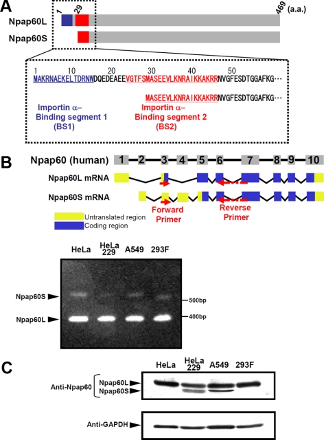
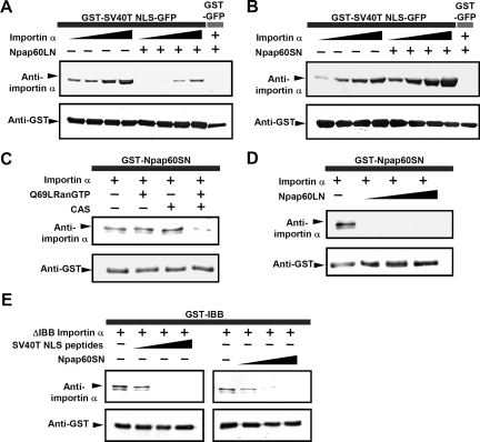
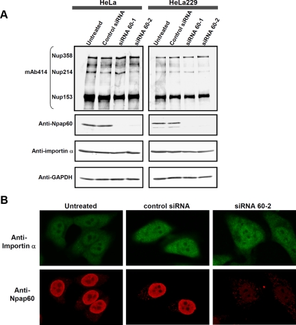
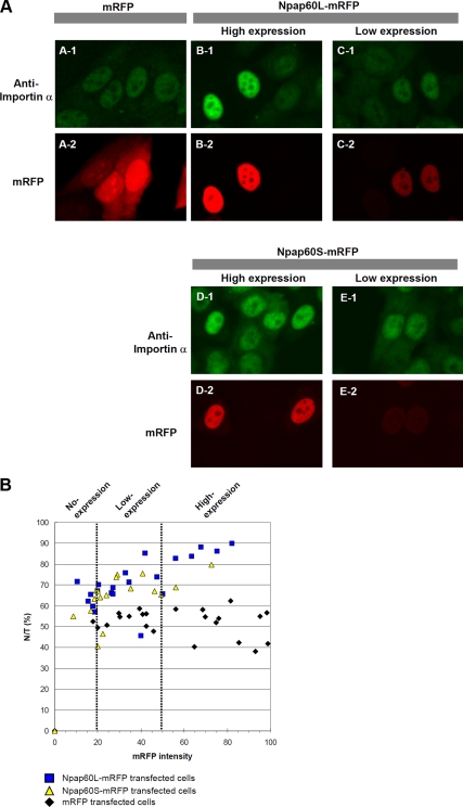
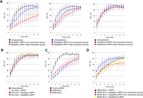
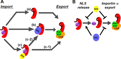
Npap60 (Nup50) is a nucleoporin that binds directly to importin alpha. In humans, there are two Npap60 isoforms: the long (Npap60L) and short (Npap60S) forms. In this study, we provide both in vitro and in vivo evidence that Npap60L and Npap60S function differently in nuclear protein import. In vitro binding assays revealed that Npap60S stabilizes the binding of importin alpha to classical NLS-cargo, whereas Npap60L promotes the release of NLS-cargo from importin alpha. In vivo time-lapse experiments showed that when the Npap60 protein level is controlled, allowing CAS to efficiently promote the dissociation of the Npap60/importin alpha complex, Npap60S and Npap60L suppress and accelerate the nuclear import of NLS-cargo, respectively. These results demonstrate that Npap60L and Npap60S have opposing functions and suggest that Npap60L and Npap60S levels must be carefully controlled for efficient nuclear import of classical NLS-cargo in humans. This study provides novel evidence that nucleoporin expression levels regulate nuclear import efficiency.


Figures

Figure 1.
Figure 1.
Expression of the Npap60 isoforms. (A) Diagrams of the Npap60 isoforms. The importin α-binding segments 1 (BS1; 1-15 aa; blue) and 2 (BS2; 23-46 aa; red) of Npap60L are indicated. BS1 is thought to be involved in releasing NLS-cargo from importin α, whereas BS2 is believed to function as a scaffold (Matsuura et al., 2005). Npap60S lacks 1-28 aa of Npap60L, which includes BS1. (B) Diagrams of the genomic structure and mRNAs of Npap60L (NM_007172,3) and Npap60S (NM_153645,2). Numbered boxes indicate the exons. The human Npap60 gene is composed of 10 exons. Yellow boxes and blue boxes indicate the untranslated regions and coding regions, respectively. Arrows indicate the locations of the RT-PCR primers. Total RNA (100 ng) from HeLa, HeLa229, A549, and 293F cells was used as a template. Bands of ∼350 and 540 base pairs corresponding to Npap60L mRNA and Npap60S mRNA, respectively, were detected in all cell lines. (C) Protein expression of the endogenous Npap60 isoforms in cultured human cells. Npap60 proteins in lysates from HeLa, HeLa229, A549, and 293F cells were examined by Western blot analysis with an anti-Npap60 antibody (1:1000). The total amount of protein was normalized to GAPDH expression in each sample. All samples were visualized with alkaline phosphatase–conjugated secondary antibodies (1:1000).
Figure 2.
Figure 2.
The Npap60 isoforms function differently in the binding between importin α and NLS-cargo. (A) Importin α alone at concentrations of 50, 100, 200 or 400 nM or in combination with 1 μM of Npap60LN was incubated with 1 μM of GST-NLS-GFP immobilized on glutathione beads, and the bound proteins were analyzed by Western blotting. (B) Importin α alone at concentrations of 50, 100, 200 or 400 nM or in combination with 1 μM Npap60SN was incubated with 1 μM immobilized GST-NLS-GFP. (C) A mixture of 50 nM of importin α and either 2 μM Q69LRan or 1 μM of CAS, or both was incubated with 500 nM of immobilized GST-Npap60SN. (D) A mixture of 50 nM importin α alone or in combination with 1, 2, or 4 μM Npap60LN was incubated with 1 μM of immobilized GST-Npap60SN. (E) ΔIBB-importin α (250 nM) and either the NLS peptide or Npap60SN were incubated with 1 μM immobilized GST-IBB. GST-fusion proteins, GFP-fusion proteins, and importin α mutants were detected using anti-GST (1:1000), anti-GFP (1:1000), and anti-karyopherin α2 (1:1000) antibodies, respectively. All samples were visualized with alkaline phosphatase–conjugated secondary antibodies (1:1000).
Figure 3.
Figure 3.
siRNA-mediated knockdown of the Npap60 isoforms does not affect the localization of endogenous importin α. (A) HeLa and HeLa229 cells were transfected with control siRNA, siRNA 60-1, or siRNA 60-2. After 24 h, whole-cell lysates were prepared. The endogenous Npap60 isoforms, importin α, Nup153, Nup214, and Nup358 were detected by Western blot analysis. The Npap60 isoforms and importin α were detected with an anti-Npap60 antibody (1:1000) and anti-karyopherin α/Rch-1 antibody (1:200), respectively. Other nucleoporins were detected with mAb 414, which recognizes FG nucleoporins, including Nup153, Nup214, and Nup358. The total protein levels were normalized to GAPDH expression in each sample. (B) HeLa cells were either untransfected or transfected with a control siRNA or siRNA 60-2. After fixing the cells, endogenous Npap60 and importin α were detected with an anti-Npap60 antibody (1:100) and anti-karyopherin α/Rch-1 antibody (1:200), followed by Alexa 568– and Alexa 488–conjugated secondary antibodies, respectively.
Figure 4.
Figure 4.
Overexpression of the Npap60 isoforms induces the nuclear accumulation of endogenous importin α. (A) HeLa cells were transfected with expression vectors encoding mRFP, Npap60L-mRFP, or Npap60S-mRFP. Twenty-four hours after transfection, the cells were fixed and examined by immunofluorescence using an anti-karyopherin α/Rch-1 antibody, followed by an Alexa 488–conjugated secondary antibody (A-1–E-1). A-2 shows cells expressing mRFP. B-2 and D-2 show cells expressing high levels of the mRFP-fused Npap60 isoforms, whereas C-2 and E-2 show cells expressing low levels of the mRFP-fused Npap60 isoforms. Importin α accumulated in nuclei depending on the expression levels of the exogenous Npap60 isoforms (B-1–E-1). (B) The nuclear-to-total ratios of endogenous importin α were plotted relative to the mRFP intensity (0–100). Based on the mRFP intensity, the cells were sorted into the no-expression group (0–20), low-expression group (20–50), or high-expression group (50–100). Black diamonds, blue squares, and yellow triangles indicate mRFP-, Npap60L-mRFP–, and Npap60S-mRFP–transfected cells, respectively.
Figure 5.
Figure 5.
In vivo time-lapse analysis. A mixture of 4 mg/ml GST-SV40T NLS-GFP and 0.5 mg/ml Alexa 647–conjugated anti-mouse IgG, as the injection marker, was injected into the cytoplasm of HeLa cells. The nuclear-to-total ratios of GST-NLS-GFP (N/T [%]) were calculated based on the intensity of GFP fluorescence at each time point. (A) Import efficiencies of cells transfected with Npap60L-mRFP, Npap60S-mRFP, or Npap60ΔN-mRFP. HeLa cells were transfected with Npap60L-mRFP (left), Npap60S-mRFP (middle), or Npap60ΔN-mRFP (right). After 24 h, time-lapse analyses were performed. The expression levels of the mRFP-fused proteins were used to classify cells into low- or high-expression groups based on the mRFP intensities (see Materials and Methods). The black line indicates the average rates of untransfected cells (n = 6). Blue and red lines indicate the rates of low-expression cells (Npap60L: n = 15, Npap60S: n = 13, Npap60ΔN: n = 5) and high-expression cells (Npap60L: n = 4, Npap60S: n = 3, Npap60ΔN: n = 3), respectively. (B) Import efficiencies of cells cotransfected with both His-tagged CAS and either mRFP (yellow line, n = 8), Npap60L-mRFP (blue line, n = 5), or Npap60S-mRFP (red line, n = 6). The black line indicates the average rates of untransfected cells. (C) Import efficiencies of Npap60-knockdown cells. HeLa cells were transfected with either siRNA 60-1 (blue line, n = 8), siRNA 60-2 (red line, n = 7), or a negative control siRNA (black line, n = 7). After 48 h, time-lapse analyses were performed. siRNA 60-1 and siRNA 60-2 target different sites that are conserved between the Npap60L and Npap60S mRNAs. (D) HeLa cells were transfected with siRNA 60-2. After 24 h, the cells were further transfected with either Npap60L-mRFP (blue line, n = 5), Npap60S-mRFP (red line, n = 4), Npap60ΔN-mRFP (yellow line, n = 3), or mRFP (black line, n = 6). After 24 h, time-lapse analyses were performed. In this assay, only the low-expression groups were included in the statistical analysis. All data in the graphs are the means ± SD.
Figure 6.
Figure 6.
Model of the nucleocytoplasmic recycling of importin α mediated by the Npap60 isoforms. (A) After importin β is released by RanGTP, importin α may be recycled via the following pathways in the nucleus. (a) Importin α releases NLS-cargo by auto-inhibition. Then, CAS/RanGTP exports cargo-free importin α. (b) Npap60L binds to the importin α/NLS-cargo complex and releases the NLS-cargo from importin α. Next, Npap60L is replaced by CAS/RanGTP. (c) Npap60S binds to importin α/NLS-cargo and stabilizes this complex. (c-1) CAS/RanGTP binds to importin α and releases Npap60S. Then, importin α releases NLS-cargo by auto-inhibition. (c-2) Npap60S is replaced by Npap60L. (B) Npap60S inhibits both the release of NLS-cargo and the export of importin α. On the other hand, Npap60L promotes the release of NLS-cargo and inhibits the export of importin α. Therefore, when the expression levels of the Npap60 isoforms are high relative to CAS, the import efficiency of NLS-cargo decreases. When the expression levels of the Npap60 isoforms are appropriate for CAS to bind importin α and release the Npap60 isoforms, Npap60L increases the import efficiency while Npap60S decreases the import efficiency.

Similar articles

Cited by

References

    1. Bayliss R., Littlewood T., Stewart M. Structural basis for the interaction between FxFG nucleoporin repeats and importin-beta in nuclear trafficking. Cell. 2000;102:99–108. - PubMed
    1. Chook Y. M., Blobel G. Karyopherins and nuclear import. Curr. Opin. Struct. Biol. 2001;11:703–715. - PubMed
    1. Cronshaw J. M., Krutchinsky A. N., Zhang W., Chait B. T., Matunis M. J. Proteomic analysis of the mammalian nuclear pore complex. J. Cell Biol. 2002;158:915–927. - PMC - PubMed
    1. Dilworth D. J., Suprapto A., Padovan J. C., Chait B. T., Wozniak R. W., Rout M. P., Aitchison J. D. Nup2p dynamically associates with the distal regions of the yeast nuclear pore complex. J. Cell Biol. 2001;153:1465–1478. - PMC - PubMed
    1. Fan F., Liu C. P., Korobova O., Heyting C., Offenberg H. H., Trump G., Arnheim N. cDNA cloning and characterization of Npap60, a novel rat nuclear pore-associated protein with an unusual subcellular localization during male germ cell differentiation. Genomics. 1997;40:444–453. - PubMed

Publication types

MeSH terms