Francisella tularensis T-cell antigen identification using humanized HLA-DR4 transgenic mice
- PMID: 20016043
- PMCID: PMC2815527
- DOI: 10.1128/CVI.00361-09
Francisella tularensis T-cell antigen identification using humanized HLA-DR4 transgenic mice
Abstract
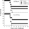
There is no licensed vaccine against the intracellular pathogen Francisella tularensis. The use of conventional mouse strains to screen protective vaccine antigens may be problematic, given the differences in the major histocompatibility complex (MHC) binding properties between murine and human antigen-presenting cells. We used engineered humanized mice that lack endogenous MHC class II alleles but that express a human HLA allele (HLA-DR4 transgenic [tg] mice) to identify potential subunit vaccine candidates. Specifically, we applied a biochemical and immunological screening approach with bioinformatics to select putative F. tularensis subsp. novicida T-cell-reactive antigens using humanized HLA-DR4 tg mice. Cell wall- and membrane-associated proteins were extracted with Triton X-114 detergent and were separated by fractionation with a Rotofor apparatus and whole-gel elution. A series of proteins were identified from fractions that stimulated antigen-specific gamma interferon (IFN-gamma) production, and these were further downselected by the use of bioinformatics and HLA-DR4 binding algorithms. We further examined the validity of this combinatorial approach with one of the identified proteins, a 19-kDa Francisella tularensis outer membrane protein (designated Francisella outer membrane protein B [FopB]; FTN_0119). FopB was shown to be a T-cell antigen by a specific IFN-gamma recall assay with purified CD4(+) T cells from F. tularensis subsp. novicida DeltaiglC-primed HLA-DR4 tg mice and cells of a human B-cell line expressing HLA-DR4 (DRB1*0401) functioning as antigen-presenting cells. Intranasal immunization of HLA-DR4 tg mice with the single antigen FopB conferred significant protection against lethal pulmonary challenge with an F. tularensis subsp. holarctica live vaccine strain. These results demonstrate the value of combining functional biochemical and immunological screening with humanized HLA-DR4 tg mice to map HLA-DR4-restricted Francisella CD4(+) T-cell epitopes.
Figures


Similar articles
-
Perforin- and granzyme-mediated cytotoxic effector functions are essential for protection against Francisella tularensis following vaccination by the defined F. tularensis subsp. novicida ΔfopC vaccine strain.Infect Immun. 2012 Jun;80(6):2177-85. doi: 10.1128/IAI.00036-12. Epub 2012 Apr 9. Infect Immun. 2012. PMID: 22493083 Free PMC article.
-
Vaccination with a defined Francisella tularensis subsp. novicida pathogenicity island mutant (DeltaiglB) induces protective immunity against homotypic and heterotypic challenge.Vaccine. 2009 Sep 18;27(41):5554-61. doi: 10.1016/j.vaccine.2009.07.034. Epub 2009 Aug 3. Vaccine. 2009. PMID: 19651173 Free PMC article.
-
Chlamydial protease-like activity factor induces protective immunity against genital chlamydial infection in transgenic mice that express the human HLA-DR4 allele.Infect Immun. 2006 Dec;74(12):6722-9. doi: 10.1128/IAI.01119-06. Epub 2006 Oct 2. Infect Immun. 2006. PMID: 17015458 Free PMC article.
-
An approach to the identification of T cell epitopes in the genomic era: application to Francisella tularensis.Immunol Res. 2009 Dec;45(2-3):218-28. doi: 10.1007/s12026-009-8103-z. Epub 2009 Feb 11. Immunol Res. 2009. PMID: 19212707 Free PMC article. Review.
-
The Brief Case: Suspicious Gram-Negative Coccobacilli-Francisella tularensis subsp. novicida Isolated from an Immunocompromised Patient.J Clin Microbiol. 2023 Jun 20;61(6):e0078722. doi: 10.1128/jcm.00787-22. Epub 2023 Jun 20. J Clin Microbiol. 2023. PMID: 37338229 Free PMC article. Review. No abstract available.
Cited by
-
Helper T cell epitope-mapping reveals MHC-peptide binding affinities that correlate with T helper cell responses to pneumococcal surface protein A.PLoS One. 2010 Feb 25;5(2):e9432. doi: 10.1371/journal.pone.0009432. PLoS One. 2010. PMID: 20195541 Free PMC article.
-
Separation of Bioactive Small Molecules, Peptides from Natural Sources and Proteins from Microbes by Preparative Isoelectric Focusing (IEF) Method.J Vis Exp. 2020 Jun 14;(160):10.3791/61101. doi: 10.3791/61101. J Vis Exp. 2020. PMID: 32597857 Free PMC article.
-
Immunity to Francisella.Front Microbiol. 2011 Feb 16;2:26. doi: 10.3389/fmicb.2011.00026. eCollection 2011. Front Microbiol. 2011. PMID: 21687418 Free PMC article.
-
Production of outer membrane vesicles and outer membrane tubes by Francisella novicida.J Bacteriol. 2013 Mar;195(6):1120-32. doi: 10.1128/JB.02007-12. Epub 2012 Dec 21. J Bacteriol. 2013. PMID: 23264574 Free PMC article.
-
Microbial co-infection alters macrophage polarization, phagosomal escape, and microbial killing.Innate Immun. 2018 Apr;24(3):152-162. doi: 10.1177/1753425918760180. Epub 2018 Feb 26. Innate Immun. 2018. PMID: 29482417 Free PMC article.
References
-
- Altschul, S. F., W. Gish, W. Miller, E. W. Myers, and D. J. Lipman. 1990. Basic local alignment search tool. J. Mol. Biol. 215:403-410. - PubMed
-
- Anthony, L. S., and P. A. Kongshavn. 1987. Experimental murine tularemia caused by Francisella tularensis, live vaccine strain: a model of acquired cellular resistance. Microb. Pathog. 2:3-14. - PubMed
-
- Bhasin, M., A. Garg, and G. P. Raghava. 2005. PSLpred: prediction of subcellular localization of bacterial proteins. Bioinformatics 21:2522-2524. - PubMed
-
- Bhasin, M., and G. P. Raghava. 2004. SVM based method for predicting HLA-DRB1*0401 binding peptides in an antigen sequence. Bioinformatics 20:421-423. - PubMed
Publication types
MeSH terms
Substances
Grants and funding
LinkOut - more resources
Full Text Sources
Research Materials
Miscellaneous

