. 2010 Jan 1;123(Pt 1):51-61.
doi: 10.1242/jcs.056424.

Integrin activation by Fam38A uses a novel mechanism of R-Ras targeting to the endoplasmic reticulum

Affiliations

Integrin activation by Fam38A uses a novel mechanism of R-Ras targeting to the endoplasmic reticulum

Brian J McHugh et al. J Cell Sci. .

Abstract

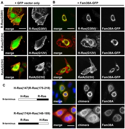
The integrin family of heterodimeric cell-surface receptors are fundamental in cell-cell and cell-matrix adhesion. Changes to either integrin-ligand affinity or integrin gene expression are central to a variety of disease processes, including inflammation, cardiovascular disease and cancer. In screening for novel activators of integrin-ligand affinity we identified the previously uncharacterised multi-transmembrane domain protein Fam38A, located at the endoplasmic reticulum (ER). siRNA knockdown of Fam38A in epithelial cells inactivates endogenous beta1 integrin, reducing cell adhesion. Fam38A mediates integrin activation by recruiting the small GTPase R-Ras to the ER, which activates the calcium-activated protease calpain by increasing Ca(2+) release from cytoplasmic stores. Fam38A-induced integrin activation is blocked by inhibition of either R-Ras or calpain activity, or by siRNA knockdown of talin, a well-described calpain substrate. This highlights a novel mechanism for integrin activation by Fam38A, utilising calpain and R-Ras signalling from the ER. These data represent the first description of a novel spatial regulator of R-Ras, of an alternative integrin activation-suppression pathway based on direct relocalisation of R-Ras to the ER, and of a mechanism linking R-Ras and calpain signalling from the ER with modulation of integrin-ligand affinity.


Figures

Fig. 1.
Fig. 1.
Screen for novel genes that rescue integrin suppression. (A) Representation of the CHO(αβ-py) screen for cDNA clones that `rescue' H-Ras(G12V)-induced integrin suppression. Left panel: transient transfection of H-Ras(G12V) inactivates integrins and reduces binding of the ligand mimetic antibody PAC-1. Representative dot blot shows PAC-1 binding versus the transfection efficiency marker Tac-α5 (black box, flow sort gate). Right panel: transient co-transfection of H-Ras(G12V) and either individual or pools of cDNAs might result in `rescue' of the H-Ras(G12V) suppression (i.e. integrin activation) allowing PAC-1 binding. Rescue might be caused via direct inhibition of H-Ras(G12V) activity, or bypassing H-Ras(G12V) activity. The corresponding flow cytometry shows corresponding cell population shift in PAC-1 binding (upper quadrants) following rescue of integrin suppression. (B) Activation indices of a selection of single cDNAs from the above screen show rescue of H-Ras(G12V)-induced suppression by cDNA clone #480.
Fig. 2.
Fig. 2.
Fam38A protein organisation and integrin activation. (A) Scheme of full-length Fam38A, the cDNA clone #480 and deletion mutants Fam38A(ΔC) and #480(ΔTM), highlighting predicted transmembrane domains. (B) Activation indices (relative to vector-alone transfection) of constructs shown in A in CHO(αβ-py) cells, without (white bars) or with (black bars) 0.5 μg H-Ras(G12V). Results show partial and complete rescue of H-Ras(G12V)-mediated integrin suppression by cDNA clone #480 and full-length Fam38A (1 μg), respectively, but not deletion constructs Fam38A(ΔC) or 480(ΔTM) (1 μg). Data shown represent mean ± s.e.m., n=3; **P<0.01.
Fig. 3.
Fig. 3.
Fam38A siRNA knockdown reduces β1-integrin affinity. (A) siRNA knockdown of Fam38A expression by oligo#3 and oligo#4, assessed by western blot and RT-PCR. β-actin is shown as loading control. (B) Relative expression of Fam38A in oligo#3- and oligo#4-treated cells compared with control siRNA, quantified by real-time PCR. (C) Flow cytometry of HeLa cells stained with β1 integrin antibodies CD29 (HUTS-21) and CD29 (K20). Histograms show native binding (white histogram) and the effect of EDTA (light grey histogram) and Mn2+ (dark grey histogram) on antibody binding, demonstrating the affinity-dependent nature of HUTS-21 binding, but not K20. (D) Activation indices of HUTS-21 binding on HeLa cells transiently transfected with Fam38A, treated with Fam38A siRNA, or transiently transfected with H-Ras(G12V) or R-Ras(G38V). (E) Flow cytometry histograms comparing HUTS-21 and K20 binding in control or Fam38A-siRNA-treated HeLa cells. GeoMean values are shown in top right corners.
Fig. 4.
Fig. 4.
Fam38A siRNA causes integrin-dependent cell detachment. (A-D) Phase contrast microscopy comparing HeLa cells treated with control oligo or Fam38A siRNA, showing cell detachment (A,B; scale bar: 30 μm) and cell morphology defects (C,D, Scale bar: 5 μm). (E-H) Confocal microscopy of Fam38A-depleted cells (E) compared with control oligo (F). Anti-paxillin-stained focal adhesions, green; rhodamine-phalloidin-labelled actin cytoskeleton, red; DAPI, blue. Corresponding paxillin-only staining is shown in G and H. Scale bar: 5 μm. (I-N) Phase contrast microscopy of control or Fam38A oligo#3-treated cells without (I,K,M) or with (J,L,N) TS2/16 integrin-activating antibody, showing rescue of adhesion defects at 72 and 96 hours after siRNA treatment. Scale bar: 20 μm.
Fig. 5.
Fig. 5.
Fam38A localises to the ER. Confocal microscopy of: (A) CHO(αβ-py) cells stained for Myc-labelled Fam38A (red), β3 integrin (green) and DAPI (blue). (B) ER tracker or SERCA2 (blue channel) co-staining with: Fam38A-GFP, #480-GFP, Myc-labelled Fam38A(ΔC), Myc-labelled #480(ΔTM) in CHO-K1 cells; and endogenous Fam38A staining in HeLa cells by anti-Fam38A antibody (green). Scale bars: 5 μm.
Fig. 6.
Fig. 6.
Calcium release and calpain activity is reduced by Fam38A depletion. (A) Representation of stimulation of Ca2+ release from the ER, resulting in increased calpain activity and subsequent downstream signalling events. Activity of calpain 1 and 2 requires the common subunit CapnS1. (B) Representative fluorimetry data plots of Fura2-AM fluorescence over time, comparing control oligo and Fam38A-siRNA-depleted cells. Ca2+ release was stimulated at 50 seconds with Thapsigargin. CaCl2 was added at 120 seconds to monitor re-uptake of external Ca2+. (C) Quantitation of Ca2+ release by control and Fam38A-depleted cells (area under the curve calculated with GraphPad Prism), shown as percentage relative to control. (D) Quantitation of calpain activity (DABCYL cleavage) in HeLa cells transfected with vector only (control), Fam38A or Fam38A siRNA; pre-incubated with calpain inhibitors PD150606 or MDL28170; or treated with CapnS1 siRNA. (E) Western blot showing equal expression of calpain 1 and 2 in control and Fam38A-siRNA-treated cell lysates (left panel), and knockdown of CapnS1 subunit expression by siRNA (right panel). Data shown represent mean ± s.e.m., n=4; P<0.05.
Fig. 7.
Fig. 7.
Fam38A recruits R-Ras to the ER. (A) Confocal microscopy of CHO-K1 cells transfected with Myc-labelled R-Ras(G38V), H-Ras(G12V) or RalA(G23V) and co-transfected with GFP vector-only. Scale bar: 5 μm. (B) CHO-K1 cells transfected as in A and co-transfected with Fam38A-GFP. Scale bar: 5 μm. (C) Confocal microscopy of CHO-K1 cells transiently co-transfected with Fam38A-GFP (green channel) and the H-Ras/R-Ras chimeras H-Ras(147)R-Ras(175-218) and R-Ras(174)H-Ras(148-189) (red channel). DAPI staining (blue) is shown in merged image. Diagrams on the left represent the proportions of H-Ras and R-Ras in each chimera. Scale bar: 5 μm.
Fig. 8.
Fig. 8.
Active R-Ras rescues calcium release and calpain activity defects in Fam38A-depleted cells. (A) Fluo-4 fluorescence quantitation in Thapsigargin-induced HeLa cells transfected with vector only (control), Fam38A, R-Ras(G38V), R-Ras(T43N), or in cells treated with Fam38A siRNA and subsequently transfected with the constructs shown. Data shown represent mean ± s.e.m., n=4; *P<0.05. (B) Quantitation of calpain 1 activity (DABCYL cleavage) in HeLa cells transfected or treated with siRNA as in A. Data shown represent mean ± s.e.m., n=4; *P<0.05. (C) R-Ras activation assay on transfected CHO-K1 lysates transfected with WT-R-Ras ± Fam38A, or ± PD150606 treatment followed by serum shock, showing binding of activated R-Ras to GST-RafRBD beads. R-Ras(G38V)-transfected lysate is shown as a positive control. (D) Confocal microscopy of transiently transfected HeLa cells showing localisation of GFP-RBD construct with or without myc-tagged Fam38A. Scale bar: 5 μm.
Fig. 9.
Fig. 9.
Fam38A-induced integrin activation requires active R-Ras, calpain and talin. (A) CHO(αβ-py) activation indices, relative to vector-alone transfection, of cells transiently transfected with H-Ras(G12V), Fam38A, R-Ras(G38V) or R-Ras(T43N). (B) CHO(σβ-py) activation indices of cells transfected with H-Ras(G12V), Fam38A, R-Ras(G38V), plus PD150606 treatment. (C) Activation indices of HUTS-21 binding on HeLa cells treated with PD150606, or with CapnS1 siRNA and subsequent transfection with Fam38A or R-Ras(G38V). (D) Activation indices of HUTS-21 binding on HeLa cells treated with talin-1 siRNA and subsequent transfection with Fam38A or R-Ras(G38V). Western blot shows siRNA-induced knockdown of intact and cleaved talin (225 kDa and 190 kDa, respectively). Data shown represent mean ± s.e.m., n=4; *P<0.05.
Fig. 10.
Fig. 10.
Proposed model of mechanism of integrin affinity modulation by Fam38A. (A) Fam38A expression relocalises R-Ras to the ER, stimulating Ca2+ release and calpain activation. A combination of calpain activity and any active R-Ras present at the PM causes increased integrin affinity, ligand binding and cell adhesion. (B) Fam38A siRNA abolishes recruitment of R-Ras to the ER, reducing Ca2+ release and calpain activity. PM-localised R-Ras remains capable of activating integrins, but the net result is reduced integrin affinity and cell detachment.

Similar articles

Cited by

References

    1. Ada-Nguema, A. S., Xenias, H., Hofman, J. M., Wiggins, C. H., Sheetz, M. P. and Keely, P. J. (2006). The small GTPase R-Ras regulates organization of actin and drives membrane protrusions through the activity of PLCepsilon. J. Cell Sci. 119, 1307-1319. - PubMed
    1. Altznauer, F., Conus, S., Cavalli, A., Folkers, G. and Simon, H. U. (2004). Calpain-1 regulates Bax and subsequent Smac-dependent caspase-3 activation in neutrophil apoptosis. J. Biol. Chem. 279, 5947-5957. - PubMed
    1. Arthur, J. S., Elce, J. S., Hegadorn, C., Williams, K. and Greer, P. A. (2000). Disruption of the murine calpain small subunit gene, Capn4: calpain is essential for embryonic development but not for cell growth and division. Mol. Cell. Biol. 20, 4474-4481. - PMC - PubMed
    1. Bhatt, A., Kaverina, I., Otey, C. and Huttenlocher, A. (2002). Regulation of focal complex composition and disass.e.m.bly by the calcium-dependent protease calpain. J. Cell Sci. 115, 3415-3425. - PubMed
    1. Carragher, N. O., Walker, S. M., Scott Carragher, L. A., Harris, F., Sawyer, T. K., Brunton, V. G., Ozanne, B. W. and Frame, M. C. (2006). Calpain 2 and Src dependence distinguishes mesenchymal and amoeboid modes of tumour cell invasion: a link to integrin function. Oncogene 25, 5726-5740. - PubMed

Publication types

MeSH terms