Comparative Study
. 2009 Dec 16;29(50):15675-83.
doi: 10.1523/JNEUROSCI.4943-09.2009.

Frontal feedback-related potentials in nonhuman primates: modulation during learning and under haloperidol

Affiliations
Comparative Study

Frontal feedback-related potentials in nonhuman primates: modulation during learning and under haloperidol

Julien Vezoli et al. J Neurosci. .

Abstract

Feedback monitoring and adaptation of performance involve a medial reward system including medial frontal cortical areas, the medial striatum, and the dopaminergic system. A considerable amount of data has been obtained on frontal surface feedback-related potentials (FRPs) in humans and on the correlate of outcome monitoring with single unit activity in monkeys. However, work is needed to bridge knowledge obtained in the two species. The present work describes FRPs in monkeys, using chronic recordings, during a trial and error task. We show that frontal FRPs are differentially sensitive to successes and failures and can be observed over long-term periods. In addition, using the dopamine antagonist haloperidol we observe a selective effect on FRP amplitude that is absent for pure sensory-related potentials. These results describe frontal dopaminergic-dependent FRPs in monkeys and corroborate a human-monkey homology for performance monitoring signals.


Figures
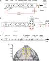
Figure 1.
Figure 1.
PST, phases of the protocol, and electrodes position. A, PST during phase 0. The task consisted in searching by trial and error the correct target and then in repeating the discovered correct response. Rectangles symbolize the successive steps in a trial and in a problem. Eye fixation and touches were controlled. See Materials and Methods for timing details. During phase zero at the touch on one target, all target items switched off and the animal either received a reward when correct or no reward when incorrect. After the outcome, another trial was initiated to proceed the search period or to enter the repetition period after the first correct choice. At the end of repetition, a visual signal (red circle) flashed on the screen to indicate the initiation of a new problem. B, PST in phase I. A visual feedback is given after a choice and during 500 ms before the actual outcome (reward or no reward). The signal to change was visually equivalent to the negative feedback. C, Experimental schedule indicating the succession of the different phases and periods of testing in time. D, Locations of transcranial electrodes reported on a stereotaxic space and drawn over a standard reconstructed brain surface fitted to a three-dimensional image (BrainMaps B3D) from http://brainmaps.org. The reference is shown in green. The two electrodes used for illustration and analyses are shown in orange.
Figure 2.
Figure 2.
RTs and saccade latencies in search and repetition periods. A, B, Average RTs in search (INC and CO1 trials) and repetition (COR trials) for monkeys S (A) and R (B). C, D, RTs in search and repetition during the successive steps and phases for the respective monkeys. E, F, Average saccade latencies in search (INC and CO1 trials) and repetition (COR trials) for monkeys S (E) and R (F).
Figure 3.
Figure 3.
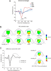
FRPs: surface maps and difference waves. A, Grand average FRPs for the two monkeys for INC, CO1, and COR trials over Phase I. The actual outcome is given at 0.5 s after feedback onset. Peak of interest is indicated. Waveforms correspond to averages between monkeys and electrodes E5/E12. B, Surface maps reconstructed for INC FRPs during Phase I for the two monkeys and at successive time points (see Materials and Methods). Each map represents the mean values for the time window used to measure ERP components. The amplitude is represented by a color code. C, Difference waves INC-COR for the two animals for the entire Phase I. On the right are represented the corresponding surface maps reconstructed at the positive peak (170 ms). Difference waves correspond to average between electrodes E5 and E12.
Figure 4.
Figure 4.
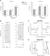
Effect of reward expectation. INC-COR and INC-CO1 difference waves. A, Grand average difference waves for INC-COR in black and INC-CO1 in gray (average between electrodes E5 and E12 between subjects). **p < 0.01, paired t test over sessions for grand average peak data. Maximum peak was measured from the time epoch represented in gray on the x-axis. B, Average amplitude of first positive peak at each step ±SEM. Statistics correspond to paired t test (*p < 0.05) between INC-CO1 and INC-COR.
Figure 5.
Figure 5.
Selective effects of haloperidol. A, Systemic haloperidol injections induced increased RT for both monkeys especially 3 h after injections. B, Values of the peak of INC-COR difference curves, for each control session (Ctr: white disks) and haloperidol sessions (30 min: measured 30 min after injection, gray disks; 3 h: measured 3 h after injection, black disks) for the two monkeys. Each disk corresponds to the average measure from a single session. The gray areas represent prediction intervals calculated from control sessions. Permutation tests showed significant differences between control and Haldol sessions at 3 h (see Results for details). Statistical significance from permutation tests: *p < 0.05; **p < 0.001; ns, nonsignificant. C, Average difference curves INC-COR for control and haloperidol (3 h) sessions for the two monkeys. D, Haloperidol had no effect on N100 potentials evoked by visual target onset. See supplemental Figure S4, available at www.jneurosci.org as supplemental material, for session per session measures.

Comment in

Similar articles

Cited by

References

    1. Amiez C, Joseph JP, Procyk E. Anterior cingulate error-related activity is modulated by predicted reward. Eur J Neurosci. 2005;21:3447–3452. - PMC - PubMed
    1. Botvinick MM, Cohen JD, Carter CS. Conflict monitoring and anterior cingulate cortex: an update. Trends Cogn Sci. 2004;8:539–546. - PubMed
    1. Brooks VB. How does the limbic system assist motor learning? a limbic comparator hypothesis. Brain Behav Evol. 1986;29:29–53. - PubMed
    1. Castner SA, Williams GV, Goldman-Rakic PS. Reversal of antipsychotic-induced working memory deficits by short-term dopamine D1 receptor stimulation. Science. 2000;287:2020–2022. - PubMed
    1. Coffin VL, Latranyi MB, Chipkin RE. Acute extrapyramidal syndrome in Cebus monkeys: development mediated by dopamine D2 but not D1 receptors. J Pharmacol Exp Ther. 1989;249:769–774. - PubMed

Publication types

LinkOut - more resources