Comparative Study
. 2009 Dec 16;29(50):15694-702.
doi: 10.1523/JNEUROSCI.3364-09.2009.

Transplanted neural precursors enhance host brain-derived myelin regeneration

Affiliations
Comparative Study

Transplanted neural precursors enhance host brain-derived myelin regeneration

Ofira Einstein et al. J Neurosci. .

Abstract

In multiple sclerosis lesions resident oligodendrocyte progenitor cells (OPCs) are present, but fail to remyelinate. In the current study we examined whether neural precursor cell (NPC) transplantation can facilitate host brain-derived remyelination. We used the chronic cuprizone-induced demyelination model in aged mice, in which slow remyelination follows cuprizone removal. NPCs were transplanted to the lateral ventricles (intracerebroventricular) of cuprizone-induced demyelinated brains. In this experimental setup, transplanted cells remained mostly in the periventricular area in an undifferentiated state. The extent of demyelination, remyelination, and proliferation of host brain regenerative cell population were examined at 1 week posttransplantation in the splenium of the corpus callosum, which was devoid of any transplanted cells. Transplantation of NPCs, but not of control, human embryonic kidney cells, significantly enhanced remyelination compared with sham-operated mice. Remyelination was performed exclusively by host brain OPCs. The proregenerative effect of transplanted NPCs was related to an increase in the proliferation of host brain OPCs. To examine the mechanism that underlies the proregenerative effect of NPCs in vitro, we used an NPC-OPC coculture system. These experiments indicated that NPCs induced the proliferation of OPCs and facilitated their differentiation into mature oligodendrocytes. The mitogenic effect of NPCs was mediated by platelet-derived growth factor-AA and fibroblast growth factor-2. In conclusion, NPC transplantation enhances host-derived myelin regeneration following chronic demyelination. This trophic effect may stimulate resident OPCs to overcome the remyelination failure in multiple sclerosis.


Figures

Figure 1.
Figure 1.
Intracerebroventricular transplantation of NPCs induces an increase in MBP content in the corpus callosum of mice after cuprizone-induced demyelination. A representative (of two experiments) Western blot (A, different bands indicate various isoforms of MBP) and densitometrical semiquantitative analysis (B) of myelin basic protein in the corpus callosum of normal (Naive) mice, after a 10 week cuprizone diet (Cup-10w), and in mice exposed to cuprizone at 2 weeks after returning to normal diet in sham-operated (Sham-2w), in NPC-transplanted (NPCTx-2w), and in HEK cell-transplanted mice (HEK-Tx-2w). MBP content was significantly reduced in response to cuprizone diet and slightly elevated following cuprizone withdrawal. NPC, but not HEK cell, transplantation induced a marked elevation in MBP content.
Figure 2.
Figure 2.
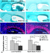
Intracerebroventricular transplantation of NPCs facilitates remyelination in the corpus callosum of mice after cuprizone-induced demyelination. Myelin content was evaluated by luxol fast blue histochemistry (A–D) and myelin basic protein immunofluorescent staining (E, F; splenium and body of the corpus callosum). Corpus callosum of a naive mouse (A, delineated by white lines), after a 10 week cuprizone diet (Cup-10w; B), and in mice exposed to cuprizone at 1 week after returning to normal diet in sham-operated (Sham-1w; C, E), and in NPC-transplanted mice (NPCTx-1w; D, F). Quantification of myelin content in the splenium (G) shows that, within 1 week of returning to normal diet, there was significant decrease in the demyelination score. In NPC-transplanted mice there was a further significant decrease in the demyelination score, compared with sham-operated mice. cc, corpus callosum; hipp, hippocampus; LV, lateral ventricle. Scale bars: (in D) A–D, 1 mm; (in F) E, F, 250 μm. Data are represented as mean ± SEM, *p < 0.05.
Figure 3.
Figure 3.
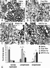
Intracerebroventricular transplantation of NPCs facilitates remyelination: high-resolution analysis in the splenium. Remyelination in the splenium of the corpus callosum was evaluated on toluidine blue-stained semithin sections of naive mice (A), after a 10 week cuprizone diet (Cup-10w; B), and in mice exposed to cuprizone at 1 week after returning to normal diet in sham-operated (Sham-1w; C) and in NPC-transplanted (NPCTx-1w; D) mice. In naive mice there were mainly normal axons (A, E), whereas after the 10 week cuprizone diet there were mainly demyelinated axons (B, E). One week after returning to normal diet, there was a significant rise in the fraction of remyelinated axons in sham-operated mice (C, E). In NPC-transplanted mice there was a significantly higher fraction of remyelinated axons (D, E). Scale bar: (in D) A–D, 5 μm. Data are represented as mean ± SEM, *p < 0.05.
Figure 4.
Figure 4.
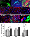
NPC transplantation enhances the repopulation of chronic cuprizone-induced demyelinated lesions by host brain-derived oligodendrocyte progenitor cells. Low-power field fluorescence microscopy (A, arrows; needle tract), followed by high-power field microscopy (B) indicated that at 1 week posttransplantation the GFP+ NPCs remained mainly in the periventricular area and did not reach the splenium of the corpus callosum. Confocal microscopy of GFP transgenic mouse brain indicated that MBP+ oligodendrocytes coexpress GFP (C; GFP, green; MBP, red; asterisk indicates location of nucleus according to dark-field microscopy). NG2+ OPCs (red) were detected in the splenium of the corpus callosum of naive mice (D) after 10 weeks of cuprizone exposure (Cup-10w; E), in mice exposed to cuprizone at 1 week after cuprizone withdrawal in sham-operated mice (Sham-1w; F), and in NPC-transplanted mice (NPCTx-1w; G). GFP+ cells had penetrated up to few cell layers in the periventricular area (H), but not further in the white matter. No GFP+ cells were observed in the splenium, where all NG2+ OPCs were host brain-derived (G). Also in the body of the corpus callosum the vast majority of OPCs were GFP− host brain cells (H, I). OPC numbers increased significantly after 10 weeks' cuprizone exposure and returned to baseline levels within 1 week after returning to normal diet in sham-operated mice (J). In NPC-transplanted mice there was a further increase in resident GFP−, NG2+ OPC number in both the body and splenium (J). Scale bars: A, 1 mm; B, 250 μm; C, I, 10 μm; D–H (in H), 100 μm. Data are represented as mean ± SEM, *p < 0.05.
Figure 5.
Figure 5.
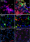
NPC transplantation enhances the proliferation of host brain-derived oligodendrocyte lineage cells in chronic cuprizone-induced demyelinated lesions. BrdU+ cells were detected (red) in the body (A, B) and in the splenium (C, D) of the corpus callosum in mice exposed to cuprizone at 1 week after cuprizone withdrawal in sham-operated (A, C) and NPC-transplanted (B, D) mice. The density of BrdU+ cells increased in the vicinity of transplanted GFP+ NPCs in the body of the corpus callosum (B), compared with sham-operated mice (A). The majority of proliferating cells were host GFP− cells (B). In the splenium, where there were no grafted cells, BrdU+ (red), NG2+ (green) cell density increased significantly in NPC-transplanted mice (D), compared with sham-operated mice (C). The density of Olig2+ cells (red) was significantly increased in the splenium of transplanted mice (G), compared with sham-operated mice (F). Scale bar: 100 μm. Data are represented as mean ± SEM, *p < 0.05.
Figure 6.
Figure 6.
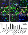
NPCs induce the proliferation of oligodendrocyte progenitor cells in vitro. Purified OPC cultures were incubated in various conditions and pulsed with BrdU to identify proliferating cells (A–I, NG2, green; BrdU, red; DAPI, blue). The total number of NG2+ cells and NG2+, BrdU+ cells per microscopic field was quantified (J). As compared with control nontreated cultures (A), the addition of PDGF-AA (B) or FGF2 (C), or both (D) significantly increased the number of proliferating OPCs (J). In cocultures, where NPCs were placed in the upper chamber of a Transwell system, a comparable increase in OPC proliferation was observed (E, J). Addition of PDGF-AA and FGF2 to the coculture had no additional effect on OPC proliferation (F, J). Addition of anti-PDGF-AA (G) or anti-FGF2 (H) neutralizing antibodies, or both antibodies (I) to the cocultures completely blocked the proliferative effect of NPCs on OPCs (J). Scale bar: A–I, 100 μm; inset in E, 10 μm. Data are represented as mean ± SEM, *p < 0.05, compared with control, nontreated OPC culture; **p < 0.05, compared with OPC–NPC coculture.
Figure 7.
Figure 7.
NPCs enhance the generation of oligodendrocytes in vitro. Purified OPC cultures were incubated in various conditions (A–E) and stained with O4 (red) and anti-GalC (green) to examine OPC differentiation. The total number of O4+ (G) or GalC+ (H) cells per microscopic field was quantified in control, nontreated cultures (A), and in FGF2-treated (B), PDGF-AA-treated (C), or FGF2 and PDGF-AA-treated (D) cultures and in OPC–NPC Transwell cocultures (E). F, High-power field showing O4+, GalC−, and O4+, GalC+ cells. FGF2 alone did not have a significant effect on the number of O4+ (G) or GalC+ (H) oligodendrocytes. PDGF-AA and the combination of PDGF-AA + FGF2 induced a 4- to 7-fold increase in the number of O4+ (G) and GalC+ (H) oligodendrocytes. Coculturing with NPCs caused a comparable effect in increasing the number of O4+ (G) and GalC+ (H) oligodendrocytes. Scale bars: A–E (in E), 100 μm; F, 10 μm. Data are represented as mean ± SEM, *p < 0.05, compared with control, nontreated culture.

Similar articles

Cited by

References

    1. Armstrong RC, Le TQ, Flint NC, Vana AC, Zhou YX. Endogenous cell repair of chronic demyelination. J Neuropathol Exp Neurol. 2006;65:245–256. - PMC - PubMed
    1. Ben-Hur T, Rogister B, Murray K, Rougon G, Dubois-Dalcq M. Growth and fate of PSA-NCAM+ precursors of the postnatal brain. J Neurosci. 1998;18:5777–5788. - PMC - PubMed
    1. Ben-Hur T, Einstein O, Mizrachi-Kol R, Ben-Menachem O, Reinhartz E, Karussis D, Abramsky O. Transplanted multipotential neural precursor cells migrate into the inflamed white matter in response to experimental autoimmune encephalomyelitis. Glia. 2003;41:73–80. - PubMed
    1. Bögler O, Wren D, Barnett SC, Land H, Noble M. Cooperation between two growth factors promotes extended self-renewal and inhibits differentiation of oligodendrocyte-type-2 astrocyte (O-2A) progenitor cells. Proc Natl Acad Sci U S A. 1990;87:6368–6372. - PMC - PubMed
    1. Calver AR, Hall AC, Yu WP, Walsh FS, Heath JK, Betsholtz C, Richardson WD. Oligodendrocyte population dynamics and the role of PDGF in vivo. Neuron. 1998;20:869–882. - PubMed

Publication types

MeSH terms