Comparative Study
. 2009 Dec 16;29(50):15819-27.
doi: 10.1523/JNEUROSCI.3776-09.2009.

Neuroprotective role of haptoglobin after intracerebral hemorrhage

Affiliations
Comparative Study

Neuroprotective role of haptoglobin after intracerebral hemorrhage

Xiurong Zhao et al. J Neurosci. .

Abstract

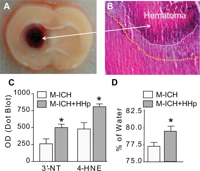
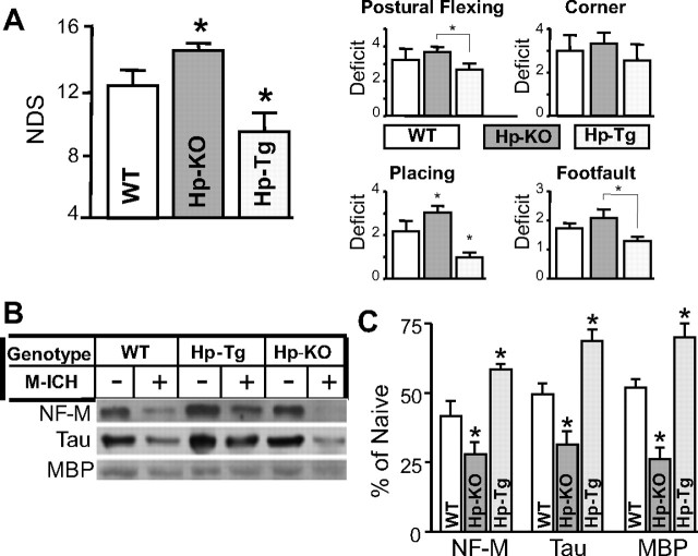
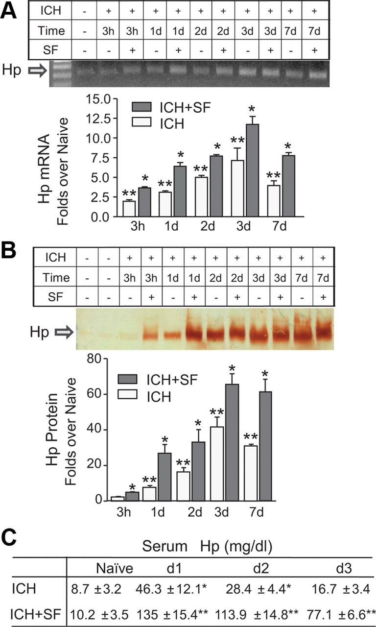
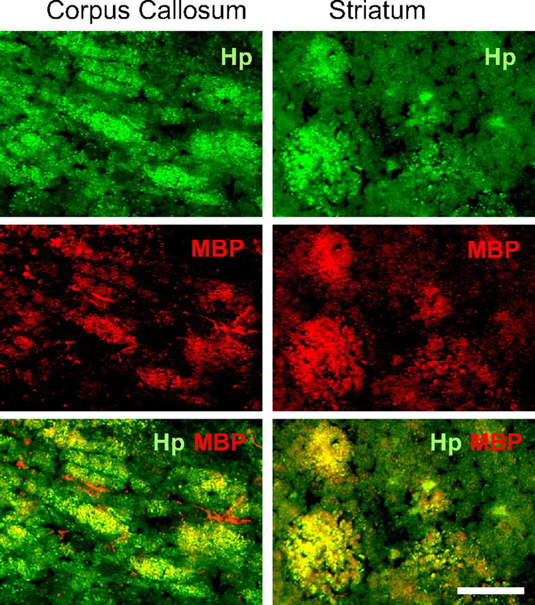
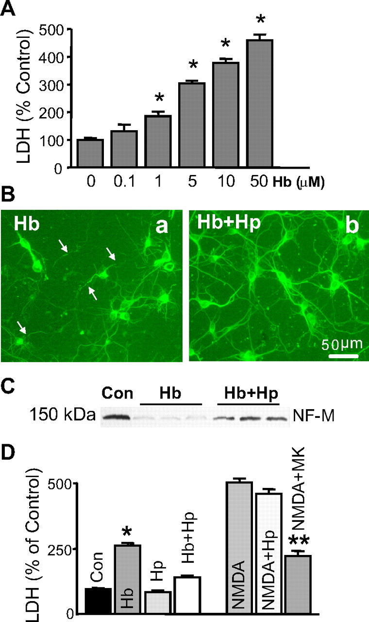
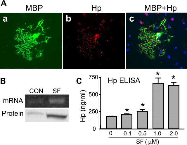
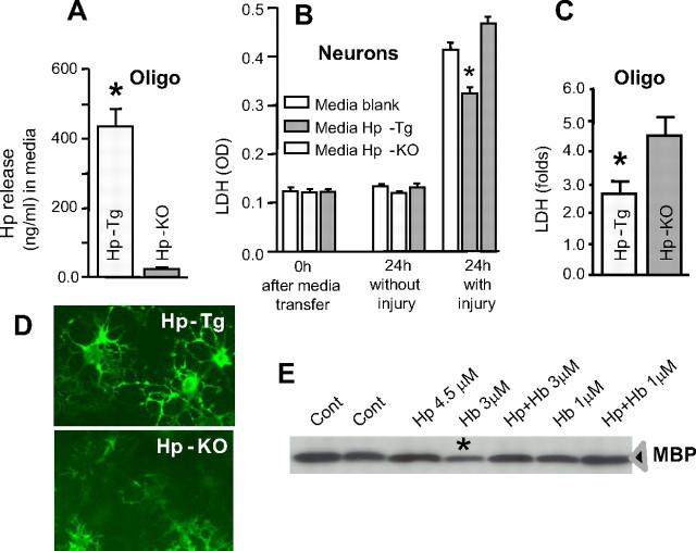
After intracerebral hemorrhage (ICH), the brain parenchyma is exposed to blood containing red blood cells (RBCs) and consequently to its lysis products. Iron-rich hemoglobin (Hb) is the most abundant protein in RBCs. When released into the brain parenchyma during hemolysis, Hb becomes a central mediator of cytotoxicity. Our study indicates that haptoglobin (Hp), an acute-phase response protein primarily synthesized in the liver and known to bind and neutralize Hb in the bloodstream, is also expressed in brain in which it plays an important role in defending neurons from damage induced by hemolytic products after ICH. We demonstrate that the Hb-induced hypohaptoglobinemia aggravates ICH-induced brain damage while pharmacologic intervention with sulforaphane to induce brain Hp is linked to a reduction in brain damage. In agreement with these findings, Hp deficiency worsens whereas Hp overexpression alleviates ICH-mediated brain injury. We also identified that oligodendroglia are the primary source of brain-derived Hp among brain cells and that oligodendroglia-released Hp plays protective roles against Hb-mediated toxicity to neurons and oligodendrocytes. We conclude that Hp, particularly the brain-derived Hp, plays cytoprotective roles and represents a potential therapeutic target for ICH treatment.


Figures

Figure 1.
Figure 1.
HHp aggravates ICH-mediated damage. A, Photograph of representative rat brain coronal section illustrating location of hematoma produced with intrastriatal injection of lysed blood (M-ICH). B, Representative photomicrograph of H&E-stained cryosection showing edema surrounding hematoma (outlined with dotted lines), as captured at 24 h after M-ICH. C, Bar graph illustrating the densitometrical analysis of immunoreactivity (immuno-dot blots technique) for 3′-NT and 4-HNE in the hematoma-affected tissue homogenates of normohaptoglobinemic control (ICH) and HHp (ICH+HHp) rats 24 h after M-ICH. The data are expressed as mean ± SEM (n = 5). *p ≤ 0.05 from the control (ICH). D, Bar graph illustrating brain edema measured by wet-to-dry weight ratio (percentage of water) at 24 h after M-ICH in the control (ICH) and HHp (ICH+HHp) rats. The data are expressed as mean ± SEM (n = 7). *p ≤ 0.05 from the control (ICH).
Figure 2.
Figure 2.
Hp protects brain from injury produced by M-ICH. A, Bar graph illustrating the composite neurological deficit scores (NDS; left panel) and deficit scores for the individual (postural reflex, corner, right forelimb placing, and right foot fault) tests at 7 d after M-ICH in WT, Hp-KO, and Hp-Tg mice. The data are expressed as mean ± SEM (n = 9). *p ≤ 0.05 from all the remaining groups. B, Photograph of immunoblot showing bands of NF-M, Tau, and MBP in hematoma-affected striatum of WT, Hp-KO, and Hp-Tg mice with (+) and without (−) M-ICH injury at 7 d after the insult. C, Bar graph of densitometrical quantification of NF-M, Tau, and MBP on the immunoblots. The data are calculated as percentage over naive animals and expressed as mean ± SEM (n = 5). *p ≤ 0.05 from all the remaining groups.
Figure 3.
Figure 3.
Hp in the hemorrhage-affected striatum is increased after ICH and is augmented by SF. The SD rats were injected intraperitoneally with either saline (ICH) or SF (ICH+SF; 5 mg/kg) at 30 min and 24 h after ICH. A, Photograph of RT-PCR products for Hp on representative agarose gel, and bar graph illustrating the densitometrical quantification of Hp mRNA at 3 h, 1, 2, 3, and 7 d after ICH. The data are calculated as mean ± SEM (n = 4) and expressed as fold increase over the naive groups. *p < 0.05 from naive and the saline control at the same time point; **p < 0.05 from naive. B, Photograph of the representative immunoblot for Hp and bar graph illustrating the densitometrical quantification of Hp protein at 3 h, 1, 2, 3, and 7 d after ICH in rats. The data are calculated as mean ± SEM (n = 4) and expressed as fold increase over the naive groups. *p < 0.05 from naive and ICH control at the same time point; **p < 0.05 from naive. C, Table indicating blood plasma Hp level in naive mice (first column) and in mice with (ICH+SF) and without (ICH) treatment with SF as determined at 1, 2, and 3 d after ICH. The data are expressed as mean ± SEM (n = 5). *p ≤ 0.05 from naive; **p < 0.05 from naive and ICH at the same time point.
Figure 4.
Figure 4.
The representative Hp expression pattern in the ICH-affected brain after ICH. The rats at 24 h after M-ICH were analyzed for presence of Hp using immunohistochemistry. Hp-immunopositive cells (green) were detected in corpus callosum and in the basal ganglia around the hematoma. The double-immunofluorescence labeling with anti-MBP (red) and anti-Hp (green) antibodies indicates that Hp colocalizes with oligodendrocytes (MBP-immunopositive cells). Scale bar, 50 μm. ICH, Hematoma.
Figure 5.
Figure 5.
A, Bar graph of LDH released into culture media by neurons in culture exposed to 0.1∼50 μm Hb for 24 h. The data are expressed as percentage over the media control and displayed as mean ± SEM (n = 3). *p ≤ 0.05 from the media control. B, Photomicrograph of MAP2 immunofluorescence in the neuronal cultures after exposure to Hb, demonstrating morphological damage. The 15-d-old neurons in culture were incubated with the following: 3 μm Hb (a) or the complexes of Hp-Hb (b) (4.5 μm Hp and 3.0 μm Hb were coincubated for 15 min before the use) for 24 h, and the morphological integrity was assessed using immunostaining for MAP2. The arrows show the breaks of neurites. C, Photograph of the immunoblot analyzing NF-M in lysates of neurons in culture subjected to Hb alone or as complexes with Hp (Hb+Hp), under experimental conditions described for B. D, Bar graph of LDH released into culture media by neurons in response to: saline (Con), 3 μm Hb, 4.5 μm Hp, Hp plus Hb, 0.5 mm NMDA, 0.5 mm NMDA plus 4.5 μm Hp, or 0.5 mm NMDA plus 10 μm MK-801 (MK). The values of LDH are expressed as percentage change over the control (Con). The data are displayed as mean ± SEM (n = 6). *p ≤ 0.05 from Con, Hp, and Hb-Hp; **p ≤ 0.05 from NMDA and NMDA plus Hp.
Figure 6.
Figure 6.
Hp is expressed by oligodendroglia and its expression is stimulated by SF. A, Photomicrograph of MBP-immunolabeled oligodendrocyte expressing Hp. The mixed neuronal–glial cocultures were incubated with 1 μm SF for 24 h and then immunolabeled for MBP (a) (green) and Hp (b) (red). c, Merged image of MBP and Hp. The nuclei were visualized with Hoechst 33258 (blue). Note that, among all cells in the field, only MBP-positive cell is immunopositive for Hp. Hp proteins are detected in the soma and fine processes of MBP-positive cells. Scale bar, 100 μm. B, Photograph of RT-PCR product for Hp on representative agarose gel (mRNA, top panel) and Hp immunoblot (protein, bottom panel), illustrating levels of Hp expression in the oligodendroglia-enriched cultures in absence (CON) or presence of SF (1 μm for 24 h). C, Bar graph quantifying Hp protein in the oligodendrocyte-enriched culture media, at 48 h after treatment with 0.1∼2 μm SF, determined by ELISA. *p < 0.05 from vehicle control.
Figure 7.
Figure 7.
A, Bar graph illustrating LDH release by 15-d-old mouse neurons in culture subjected to “ICH-like” injury (lysed RBC plus hypoxia) in the presence of conditioned media from OEC prepared from either Hp-KO or Hp-Tg mice or blank (control) media. The conditioned or blank medium was transferred into the neuronal culture (replacing two-thirds volume of the media used to grow neurons) and incubated for 15 min before induction of ICH-like injury. The cytotoxicity level after media transfer (before injury; 0 h) and at 24 h with and without induction of injury was assessed by the analysis of LDH released into the culture media. The LDH release is expressed as optical density. The data are calculated as mean ± SEM (n = 3). *p ≤ 0.05 from Hp-KO. B, Bar graph of LDH release by Hp-deficient (Hp-KO) and Hp-overexpressing (Hp-Tg) OEC on injury induced by exposure to RBC lysates. The levels of LDH released into culture media during first 16 h after induction of the insult are expressed as fold increase over the control (without RBC lysate). The data are calculated as mean ± SEM (n = 6). *p ≤ 0.05 from Hp-KO. C, Representative photomicrograph of OEC from Hp-KO or Hp-Tg mice that were exposed to RBC lysates for 16 h. Oligodendroglia were visualized by immunostaining for MBP. Note that the oligodendrocytes overexpressing Hp (Hp-Tg) show better-preserved morphology, compared with Hp-deficient (Hp-KO) ones. Scale bar, 50 μm. D, Representative Western blot for MBP as determined in homogenates from OEC exposed to 1 or 3 μm Hb in the presence or absence of 4.5 μm Hp for 24 h. *p ≤ 0.05 from control (Cont).

Similar articles

Cited by

References

    1. Allison AC, Rees WA. The binding of haemoglobin by plasma proteins (haptoglobins); its bearing on the renal threshold for haemoglobin and the aetiology of haemoglobinuria. Br Med J. 1957;2:1137–1143. - PMC - PubMed
    1. Aronowski J, Hall CE. New horizons for primary intracerebral hemorrhage treatment: experience from preclinical studies. Neurol Res. 2005;27:268–279. - PubMed
    1. Baumann H, Muller-Eberhard U. Synthesis of hemopexin and cysteine protease inhibitor is coordinately regulated by HSF-II and interferon-beta 2 in rat hepatoma cells. Biochem Biophys Res Commun. 1987;146:1218–1228. - PubMed
    1. Bhasin RR, Xi G, Hua Y, Keep RF, Hoff JT. Experimental intracerebral hemorrhage: effect of lysed erythrocytes on brain edema and blood-brain barrier permeability. Acta Neurochir Suppl. 2002;81:249–251. - PubMed
    1. Borsody M, Burke A, Coplin W, Miller-Lotan R, Levy A. Haptoglobin and the development of cerebral artery vasospasm after subarachnoid hemorrhage. Neurology. 2006;66:634–640. - PubMed

Publication types