Comparative Study
. 2009 Dec 16;29(50):15888-97.
doi: 10.1523/JNEUROSCI.4053-09.2009.

An intrinsic neuronal oscillator underlies dopaminergic neuron bursting

Affiliations
Comparative Study

An intrinsic neuronal oscillator underlies dopaminergic neuron bursting

Christopher A Deister et al. J Neurosci. .

Abstract

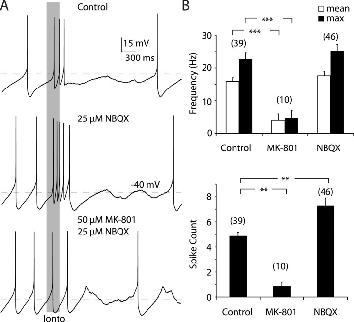
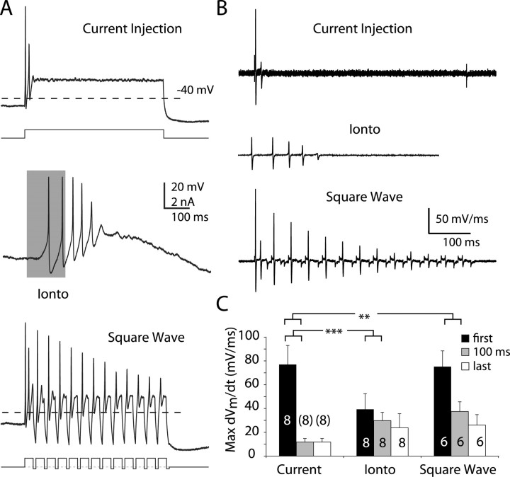
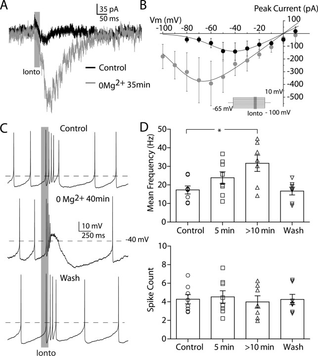
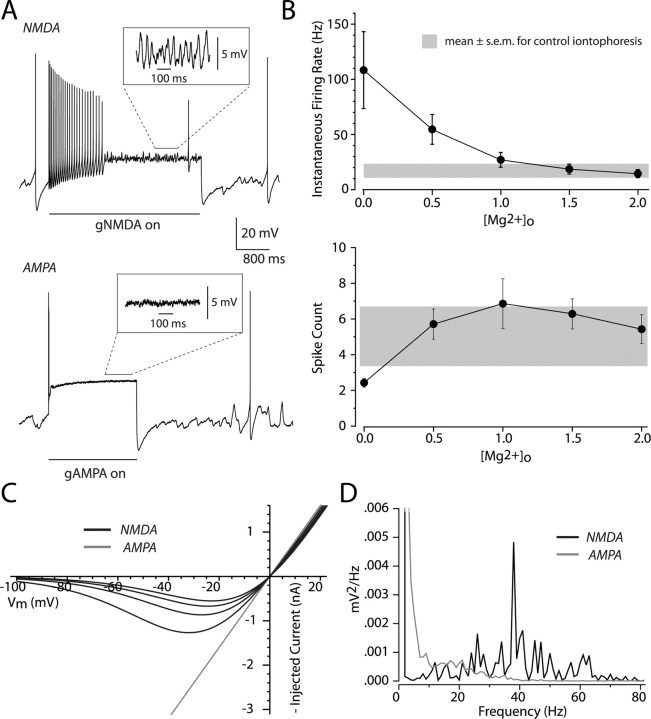
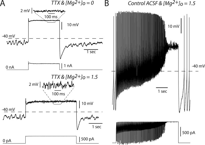
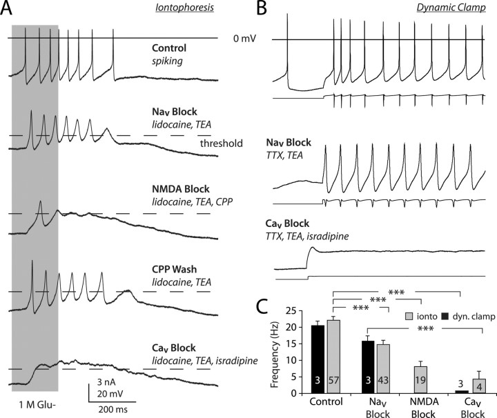
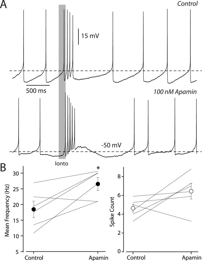
Dopaminergic neurons of the ventral midbrain fire high-frequency bursts when animals are presented with unexpected rewards, or stimuli that predict reward. To identify the afferents that can initiate bursting and establish therapeutic strategies for diseases affected by altered bursting, a mechanistic understanding of bursting is essential. Our results show that bursting is initiated by a specific interaction between the voltage sensitivity of NMDA receptors and voltage-gated ion channels that results in the activation of an intrinsic, action potential-independent, high-frequency membrane potential oscillation. We further show that the NMDA receptor is uniquely suited for this because of the rapid kinetics and voltage dependence imparted to it by Mg(2+) ion block and unblock. This mechanism explains the discrete nature of bursting in dopaminergic cells and demonstrates how synaptic signals may be reshaped by local intrinsic properties of a neuron before influencing action potential generation.


Figures

Figure 1.
Figure 1.
NMDA receptor activation is necessary for the generation of high-frequency burst firing. A, Example traces from the same neuron recorded in the whole-cell configuration. The gray bar illustrates the onset and offset of the iontophoretic application of glutamate. Top, Application of glutamate consistently led to a high-frequency episode of firing that was usually followed by brief period of quiescence, in control conditions. Middle, Application of NBQX did not block high-frequency bursting and in some cases increased the number of spikes associated with it. Bottom, Application of MK-801 consistently blocked high-frequency firing, such that the spiking associated with the iontophoresis was similar to the background firing. Note that there appears to be a slight variation of background firing rate between each of the trials; this is due to natural variability in pacemaking and was not significant. In this example, NBQX was still present when MK-801 was applied. B, MK-801 decreased the mean (top, white bars) and maximum (max) (top, black bars) intraburst firing rates and the number of spikes/burst (bottom). NBQX increased the number of spikes occurring within a burst. **p < 0.01, ***p < 0.001, one-way ANOVA plus Dunnett's test. The number above each bar is the number of cells in the sample.
Figure 2.
Figure 2.
Oscillatory inputs and glutamate iontophoresis counteract depolarization block. A, Example traces from the same neuron recorded in the whole-cell configuration. Top, The neuron was injected with a 500 ms, 700 pA current injection, to which the neuron rapidly entered a nonspiking depolarized state (Current Injection). Middle, The neuron fired at a rate of ∼25 Hz in response to a 200-ms-duration iontophoretic application of glutamate (Ionto), as indicated by the gray bar. Bottom, The neuron fired repetitively in response to a 25 Hz train of 35 ms, 700 pA current pulses (Square Wave). Note that the pulses are interleaved with 10 ms hyperpolarizing current pulses. B, Temporal derivatives (dV m/dt) of the voltage traces obtained from each protocol in A. C, Maximum dV m/dt values, for each protocol, across our sample. Black bars indicate the values for the first spike, whereas gray corresponds to the first spike after a 100 ms latency and white represents the last spike. **p < 0.01, ***p < 0.001, two-way ANOVA plus Dunnett's test. The number with each bar is the number of cells in the sample.
Figure 3.
Figure 3.
Burst firing is augmented by increasing the NMDA-mediated inward current. A, Mg2+ removal increased inward currents elicited by glutamate iontophoresis (Ionto; gray). B, Mg2+ dependence of the NMDA receptor I–V curve (average of 3 neurons recorded in whole-cell configuration). Inset, Protocol used to establish I–V curve. Glutamate was iontophoresed (25 ms, gray bar) once the cell reached steady state. Removal of Mg2+ shifts the negative slope conductance region leftward and increases the peak inward current. The I–V relationship never becomes linear. C, Example traces from the same neuron recorded in the whole-cell configuration. Top, Spiking elicited in control media with glutamate iontophoresis, indicated by gray bar. Middle, The same iontophoresis evokes a higher-frequency burst after washing in a solution that did not contain any Mg2+ for 40 min. Bottom, Washing back in control media restores normal burst firing. D, Bar charts summarizing the effect of washing solutions containing 0 Mg2+ for either 5 or >10 min, on both the mean burst firing rate (top) and the total number of spikes within the burst (bottom), in our sample.
Figure 4.
Figure 4.
The NMDA receptor's voltage dependence is a critical element in burst firing. A, An NMDA or AMPA conductance was applied to the soma of dopaminergic cells recorded in the whole-cell configuration. The conductances were modeled using a formalism for the NMDA receptor by Jahr and Stevens (1990). We treated the external Mg2+ concentration, [Mg2+]o, as a free parameter and kept the maximal conductance fixed within an experiment. Top, Example trace evoked by turning on a conductance with [Mg2+]o = 1.5 mm (NMDA). Note the presence of prominent oscillations throughout the trace even when spiking has stopped. Bottom, Trace from the same cell with [Mg2+]o = 0.0 mm (AMPA). Note the rapid decrement of spiking and lack of oscillations. B, Top, Plot of the average instantaneous firing rate as a function of [Mg2+]o for a sample of six cells. The average rate of burst firing (±SEM) evoked with iontophoresis (see Fig. 1) is shown in gray as a reference. Bottom, Plot of the number of spikes counted within 1 s. of the conductance step for the same six cells; again, the distribution of iontophoresis experiments is shown in gray. C, I–V curves for our modeled conductance calculated for different [Mg2+]o. Black traces are for [Mg2+]o = 2.0, 1.5, 1.0, and 0.5 mm (NMDA) from the smallest to largest curves, respectively. Gray trace is for [Mg2+]o = 0.0 mm (AMPA). D, Power spectra after spiking has ceased from the example traces shown in A.
Figure 5.
Figure 5.
NMDA, but not AMPA, conductances engage a high-frequency, action potential-independent oscillation. A, Dynamic clamp experiments performed in the presence of TTX and recorded in the whole-cell configuration. Top, Example electrical trace obtained for [Mg2+]o = 0.0 mm with the injected current below. Just before the conductance step, a low-frequency oscillation can be seen. During the conductance step the membrane voltage remains flat. Bottom, Same as top except [Mg2+]o = 1.5 mm. During the conductance step a higher-frequency oscillation (relative to that before the step) is observed. Below each voltage trace is the injected current waveform. B, Control firing evoked by applying the same NMDA conductance step ([Mg2+]o = 1.5) as in A, before applying TTX.
Figure 6.
Figure 6.
NMDA receptor activation triggers an intrinsic, action potential-independent, voltage oscillation. A, Iontophoresis of glutamate (gray bar) elicited a burst of action potentials from a cell recorded in the whole-cell configuration. Blockade of sodium channels (Nav block) revealed large voltage oscillations that were attenuated by NMDA and calcium channel antagonists (NMDA and Cav block, respectively). Note that all examples are from the same cell with experiments done sequentially. Dashed line indicates spike threshold. B, Same experiment as in A, except dynamic clamp was used to mimic NMDA receptor conductance. Below each voltage trace is the waveform of the injected current. C, Bar chart showing the frequency of glutamate-elicited oscillations before and after Nav block, NMDA block, or Cav block in iontophoresis (ionto) experiments (gray) and dynamic clamp (dyn. clamp) experiments (black). The number with each bar is the number of cells in the sample. ***p < 0.001, Kruskal–Wallis test (score = 42.01, p < 0.0001) plus Dunnett's test. TEA, Tetraethylammonium.
Figure 7.
Figure 7.
Burst firing is augmented, not suppressed, by SK channel blockade. A, Example traces from the same neuron recorded in the whole-cell configuration. Top, Spiking elicited in control media with glutamate iontophoresis (Ionto), indicated by gray bar. Bottom, Under the same conditions except with the addition of 100 nm apamin. Now the cell responds with a much higher frequency and more spikes. B, Summary data of the effect of 100 nm apamin, on both the mean burst firing rate (left) and the total number of spikes within the burst (right), in our sample. Each cell's values before and after drug application are indicated by individual joined lines, and the mean and SEM of our sample are given by the closed and filled circles. Asterisk indicates statistical significance (p < 0.05; paired t test).

References

    1. Amini B, Clark JW, Jr, Canavier CC. Calcium dynamics underlying pacemaker-like and burst firing oscillations in midbrain dopaminergic neurons: a computational study. J Neurophysiol. 1999;82:2249–2261. - PubMed
    1. Blythe SN, Atherton JF, Bevan MD. Synaptic activation of dendritic AMPA and NMDA receptors generates transient high-frequency firing in substantia nigra dopamine neurons in vitro. J Neurophysiol. 2007;97:2837–2850. - PubMed
    1. Brothwell SL, Barber JL, Monaghan DT, Jane DE, Gibb AJ, Jones S. NR2B- and NR2D-containing synaptic NMDA receptors in developing rat substantia nigra pars compacta dopaminergic neurones. J Physiol. 2008;586:739–750. - PMC - PubMed
    1. Calabresi P, Lacey MG, North RA. Nicotinic excitation of rat ventral tegmental neurones in vitro studied by intracellular recording. Br J Pharmacol. 1989;98:135–140. - PMC - PubMed
    1. Canavier CC. Sodium dynamics underlying burst firing and putative mechanisms for the regulation of the firing pattern in midbrain dopamine neurons: a computational approach. J Comput Neurosci. 1999;6:49–69. - PubMed

Publication types