

Intranasal CpG therapy attenuated experimental fungal asthma in a TLR9-dependent and -independent manner
- PMID: 20016192
- PMCID: PMC2883833
- DOI: 10.1159/000265531
Intranasal CpG therapy attenuated experimental fungal asthma in a TLR9-dependent and -independent manner
Abstract
Background: CpG administration abolishes airway inflammation and remodeling in acute models of allergic airway disease.
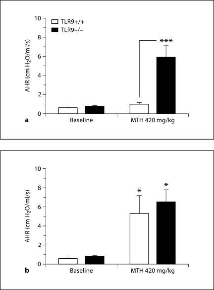
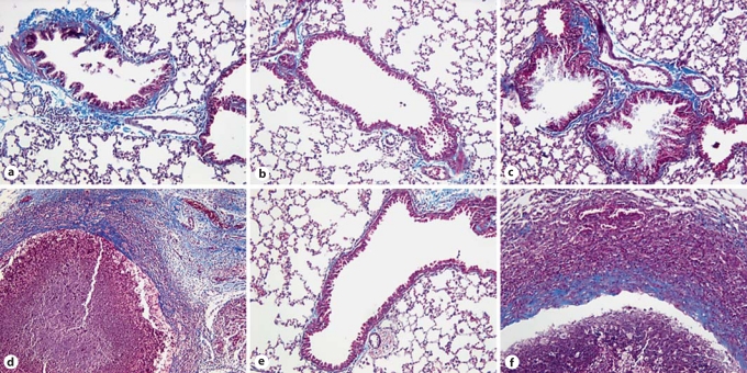
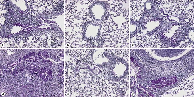
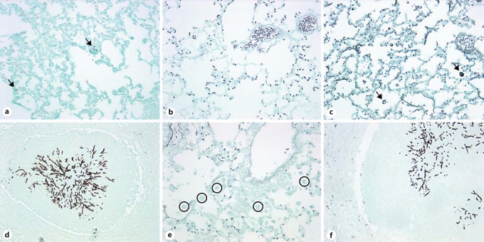
Methods: Herein, we investigated the therapeutic effect of CpG in a chronic fungal model of asthma. TLR9+/+ and TLR9-/- mice were sensitized to soluble Aspergillus fumigatus antigens and challenged with live A. fumigatus conidia. Mice were treated with intraperitoneal (IP) or intranasal (IN) CpG, or left untreated 14-28 days after conidium challenge. All features of allergic airway disease were attenuated in TLR9+/+ mice treated with IN CpG, including airway hyperresponsiveness (AHR), mucus production, and peribronchial fibrosis.
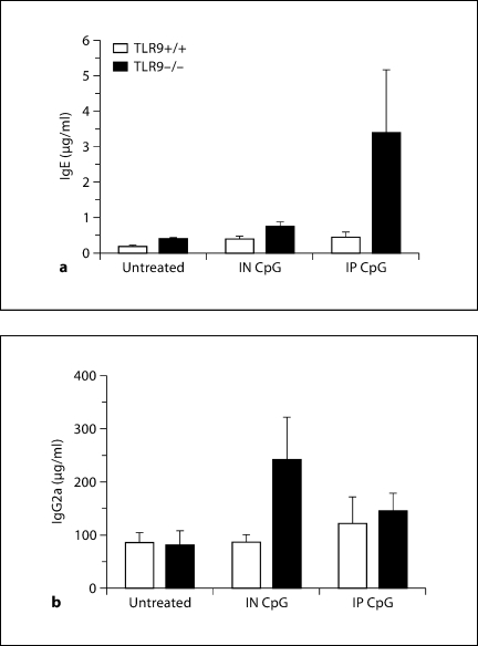
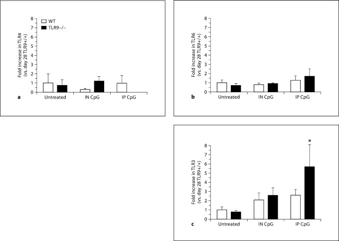
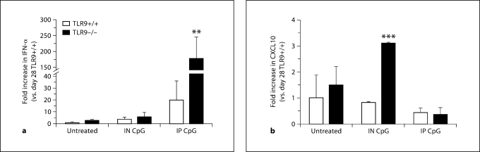
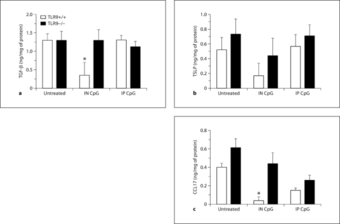
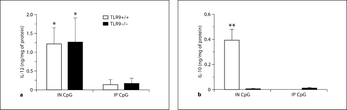
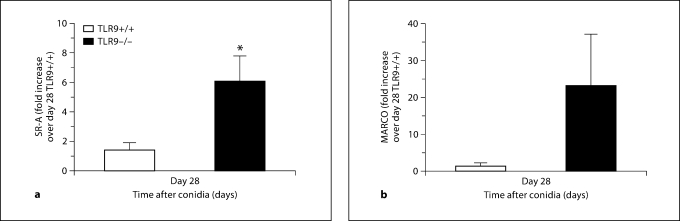
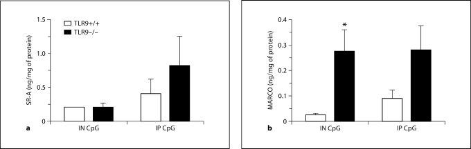
Results: TLR9-/- mice treated with IN CpG exhibited attenuated airway remodeling but not AHR. Whole-lung IL-12 levels were significantly elevated in both TLR9+/+ and TLR9-/- mice receiving IN CpG but not in either group receiving IP CpG. Whole-lung IL-10 levels were significantly elevated in IN CpG-treated TLR9+/+ mice but not in TLR9-/- mice receiving IN CpG. Increased whole-lung transcript and protein levels of the scavenger receptors SR-A and MARCO were observed in TLR9-/- mice compared with TLR9+/+ mice, possibly accounting for the CpG responsiveness in the knockout group.
Conclusions: Together, these data show that IN CpG has a therapeutic effect during established fungal asthma, which is TLR9 dependent and independent.
Copyright 2009 S. Karger AG, Basel.
Figures
Similar articles
-
Targeting ST2L potentiates CpG-mediated therapeutic effects in a chronic fungal asthma model.Am J Pathol. 2011 Jul;179(1):104-15. doi: 10.1016/j.ajpath.2011.03.032. Epub 2011 May 14. Am J Pathol. 2011. PMID: 21640974 Free PMC article.
-
Toll-like receptor 9 modulates immune responses to Aspergillus fumigatus conidia in immunodeficient and allergic mice.Infect Immun. 2009 Jan;77(1):108-19. doi: 10.1128/IAI.00998-08. Epub 2008 Oct 20. Infect Immun. 2009. PMID: 18936185 Free PMC article.
-
Intranasal administration of CpG oligodeoxynucleotides reduces lower airway inflammation in a murine model of combined allergic rhinitis and asthma syndrome.Int Immunopharmacol. 2015 Sep;28(1):390-8. doi: 10.1016/j.intimp.2015.06.028. Epub 2015 Jul 9. Int Immunopharmacol. 2015. PMID: 26163938
-
CpG oligodeoxynucleotides as TLR9 agonists: therapeutic application in allergy and asthma.BioDrugs. 2010 Aug 1;24(4):225-35. doi: 10.2165/11536140-000000000-00000. BioDrugs. 2010. PMID: 20623989 Review.
-
Toll-like receptor 9 activation with CpG oligodeoxynucleotides for asthma therapy.Drug News Perspect. 2008 Oct;21(8):434-9. doi: 10.1358/dnp.2008.21.8.1272133. Drug News Perspect. 2008. PMID: 19034349 Review.
Cited by
-
Immune responses to airborne fungi and non-invasive airway diseases.Semin Immunopathol. 2015 Mar;37(2):83-96. doi: 10.1007/s00281-014-0471-3. Epub 2014 Dec 13. Semin Immunopathol. 2015. PMID: 25502371 Review.
-
The cell biology of the innate immune response to Aspergillus fumigatus.Ann N Y Acad Sci. 2012 Dec;1273(1):78-84. doi: 10.1111/j.1749-6632.2012.06837.x. Ann N Y Acad Sci. 2012. PMID: 23230841 Free PMC article.
-
Animal Models of Aspergillosis.Comp Med. 2018 Apr 2;68(2):109-123. Comp Med. 2018. PMID: 29663936 Free PMC article. Review.
-
Modeling DNA damage-induced pneumopathy in mice: insight from danger signaling cascades.Radiat Oncol. 2017 Aug 24;12(1):142. doi: 10.1186/s13014-017-0865-1. Radiat Oncol. 2017. PMID: 28836991 Free PMC article. Review.
-
Toll-like receptor-mediated immune imbalance in asthma: controversies, breakthroughs, and future directions.Front Immunol. 2025 Jul 2;16:1605185. doi: 10.3389/fimmu.2025.1605185. eCollection 2025. Front Immunol. 2025. PMID: 40672946 Free PMC article. Review.
References
-
- Benayoun L, Druilhe A, Dombret MC, Aubier M, Pretolani M. Airway structural alterations selectively associated with severe asthma. Am J Respir Crit Care Med. 2003;167:1360–1368. - PubMed
-
- Jeffery PK. Remodeling and inflammation of bronchi in asthma and chronic obstructive pulmonary disease. Proc Am Thorac Soc. 2004;1:176–183. - PubMed
-
- Kline JNK, Arthur M. Cpg oligodeoxynucleotides. New Drugs for Asthma, Allergy and COPD. In: Hansel TT, Barnes PJ, editors. Prog Respir Res. vol 31. Basel: Karger; 2001. pp. 229–232.
-
- Racila DM, Kline JN. Perspectives in asthma: Molecular use of microbial products in asthma prevention and treatment. J Allergy Clin Immunol. 2005;116:1202–1205. - PubMed
Publication types
MeSH terms
Substances
Grants and funding
LinkOut - more resources
Full Text Sources
Medical
Research Materials
Miscellaneous
