. 2010;47(4):277-86.
doi: 10.1159/000265562. Epub 2009 Dec 16.

Site-specific connexin phosphorylation is associated with reduced heterocellular communication between smooth muscle and endothelium

Affiliations

Site-specific connexin phosphorylation is associated with reduced heterocellular communication between smooth muscle and endothelium

Adam C Straub et al. J Vasc Res. 2010.

Abstract

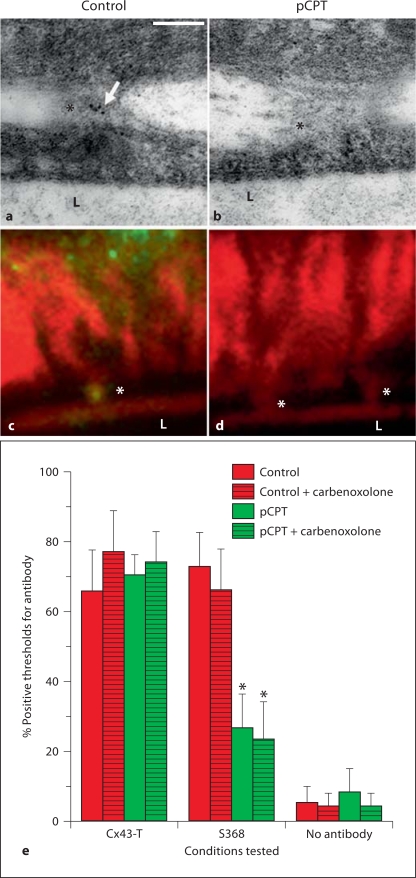
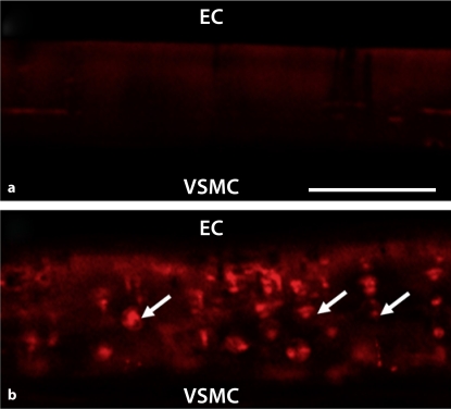
Background/aims: Myoendothelial junctions (MEJs) represent a specialized signaling domain between vascular smooth muscle cells (VSMC) and endothelial cells (EC). The functional consequences of phosphorylation state of the connexins (Cx) at the MEJ have not been explored.

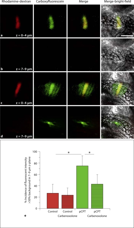
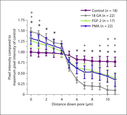
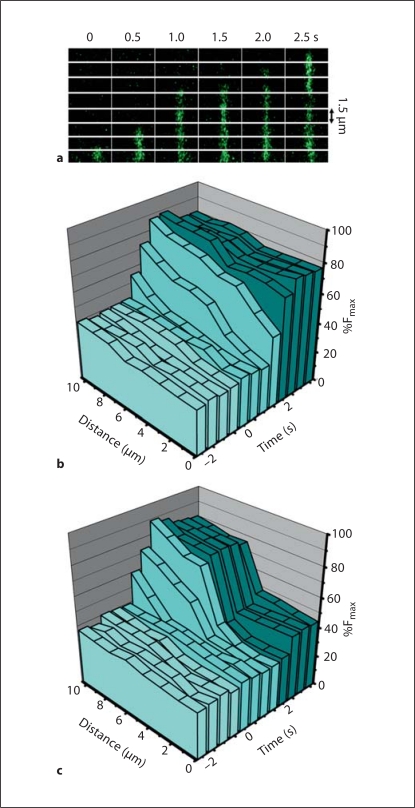
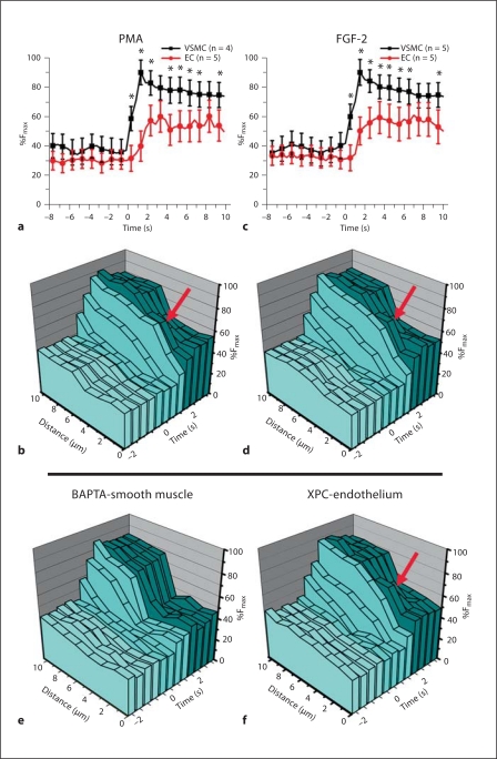
Methods/results: Application of adenosine 3',5'-cyclic monophosphate sodium (pCPT) to mouse cremasteric arterioles reduces the detection of connexin 43 (Cx43) phosphorylated at its carboxyl terminal serine 368 site (S368) at the MEJ in vivo. After single-cell microinjection of a VSMC in mouse cremaster arterioles, only in the presence of pCPT was dye transfer to EC observed. We used a vascular cell co-culture (VCCC) and applied the phorbol ester 12-O-tetradecanoylphorbol 13-acetate (PMA) or fibroblast growth factor-2 (FGF-2) to induce phosphorylation of Cx43 S368. This phosphorylation event was associated with a significant reduction in dye transfer and calcium communication. Using a novel method to monitor increases in intracellular calcium across the in vitro MEJ, we noted that PMA and FGF-2 both inhibited movement of inositol 1,4,5-triphosphate (IP(3)), but to a lesser extent Ca(2+).

Conclusion: These data indicate that site-specific connexin phosphorylation at the MEJ can potentially regulate the movement of solutes between EC and VSMC in the vessel wall.


Figures

Fig. 1
Fig. 1
Application of pCPT reduces phosphorylation of Cx43 at S368 on MEJs and actin bridges. Immunogold labeling (a, b) and actin bridge labeling (c, d) of S368 (green)/phalloidin (red) in control and pCPT-treated cremasteric arterioles. e Histogram of percent number of time that a protein was detected by antibodies on an actin bridge in mouse cremasteric arterioles. Bars are standard deviation. In a, arrow indicates gold beads, in a–d, L indicates lumen of vessel and asterisks indicate MEJs. In e, asterisks represent a significant difference between control and treatment.
Fig. 2
Fig. 2
In mouse cremaster arterioles, dye transfer from VSMC to EC is only observed after treatment with pCPT. In images a–d, raw data from single-cell microinjection of VSMC in control conditions (a, b) or conditions where pCPT was applied to the cremaster (c, d) are demonstrated. In a and c, a single VSMC is seen after microinjection with rhodamine-dextran (red) and carboxyfluoresceine (green). At least 12 VSMC were injected per experimental paradigm in 3 separate mice. The 2 images are also seen merged and superimposed over the bright-field image of the vessel. In b and d, a lower focal plane is used to observe EC. The scale bar in a is 30 μm and is representative for all images. Histogram in e is the percent incidence that the carboxyfluoresceine is observed in EC. * Significant difference between tested conditions.
Fig. 3
Fig. 3
Phosphorylation of Cx43 at S368 at the in vitro MEJ after application of PMA. Transverse view of VCCC stained for Cx43 S368 (red) in control (a) or after application of PMA (b); n = 5 different VCCC. Arrows indicate S368 labeling on the MEJ. The scale bar in a is 10 μm and is representative for all images.
Fig. 4
Fig. 4
Reduced dye transfer from smooth muscle to endothelium after treatment with FGF-2 or PMA. Biocytin was loaded into VSMC. Transfer of biocytin through the Transwell insert pores to EC at 1-μm intervals was determined by streptavidin pixel intensity. Four different experimental paradigms were tested; biocytin transfer from VSMC to EC (control, purple), biocytin transfer after application of 18 GA (18 GA, gray), biocytin transfer after application of FGF-2 (FGF-2, green) and biocytin transfer after application of PMA (PMA, blue). n is the number of pores examined per paradigm. Symbols represent p < 0.05 when compared to control at each point along the pore length: * 18 GA; ° FGF-2; + PMA.
Fig. 5
Fig. 5
Movement of an increase in intracellular calcium from VSMC to EC through the in vitro myoendothelial junction. a Line scans of a single pore from VSMC to EC over time with Fluo-4 in green. b, c The %Fmax, time and distance through the pores of the Transwell are plotted in three-dimensional walls. In control conditions, there is a spread of an increase in calcium that moves from the VSMC to the EC through the pores (b). However, when the gap junction inhibitor 18 GA is applied to the cremasteric VCCC, the increase in calcium initiated in the VSMC is halted approximately half-way through the pore – the approximate location where EC and VSMC make contact [16] (c). Application of PE occurs at time point 0 s; VSMC side of the pore is at distance 10 μm and EC side of the pore is at distance 0 μm.
Fig. 6
Fig. 6
Phosphorylation of Cx43 at S368 inhibits IP3 flux from VSMC to EC. The VCCC was treated with PMA (a, b) or FGF-2 (c, d). In a and c, measurements of the intracellular calcium in the EC and VSMC monolayers (as previously described [7]) were compared to the calcium communication moving through the in vitro MEJ in the pores of the Transwell insert (b, d). With both treatments the red arrows indicate a transient increase in calcium on the EC side of the in vitro MEJ. When VSMC were additionally loaded with BAPTA-AM (e), the transient increase in calcium on the EC side of the in vitro MEJ is no longer present, while treatment of the EC monolayer with XPC failed to inhibit the transient increase in EC calcium (f).

Similar articles

Cited by

References

    1. de Wit C, Boettcher M, Schmidt VJ. Signaling across myoendothelial gap junctions –fact or fiction? Cell Commun Adhes. 2008;15:231–245. - PubMed
    1. Sandow SL, Haddock RE, Hill CE, Chadha PS, Kerr PM, Welsh DG, Plane F. What's where and why at a vascular myoendothelial microdomain signalling complex. Clin Exp Pharmacol Physiol. 2009;36:67–76. - PubMed
    1. Heberlein K, Straub AC, Isakson BE. The myoendothelial junction: breaking through the matrix? Microcirculation. 2009;16:307–322. - PMC - PubMed
    1. Rhodin JA. The ultrastructure of mammalian arterioles and precapillary sphincters. J Ultrastruct Res. 1967;18:181–223. - PubMed
    1. Mather S, Dora KA, Sandow SL, Winter P, Garland CJ. Rapid endothelial cell-selective loading of connexin 40 antibody blocks endothelium-derived hyperpolarizing factor dilation in rat small mesenteric arteries. Circ Res. 2005;97:399–407. - PubMed

Publication types

MeSH terms