. 2010 Jan 7;463(7277):93-7.
doi: 10.1038/nature08644. Epub 2009 Dec 16.

Identification of sister chromatids by DNA template strand sequences

Affiliations

Identification of sister chromatids by DNA template strand sequences

Ester Falconer et al. Nature. .

Abstract

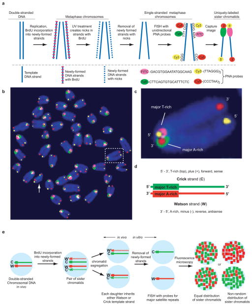
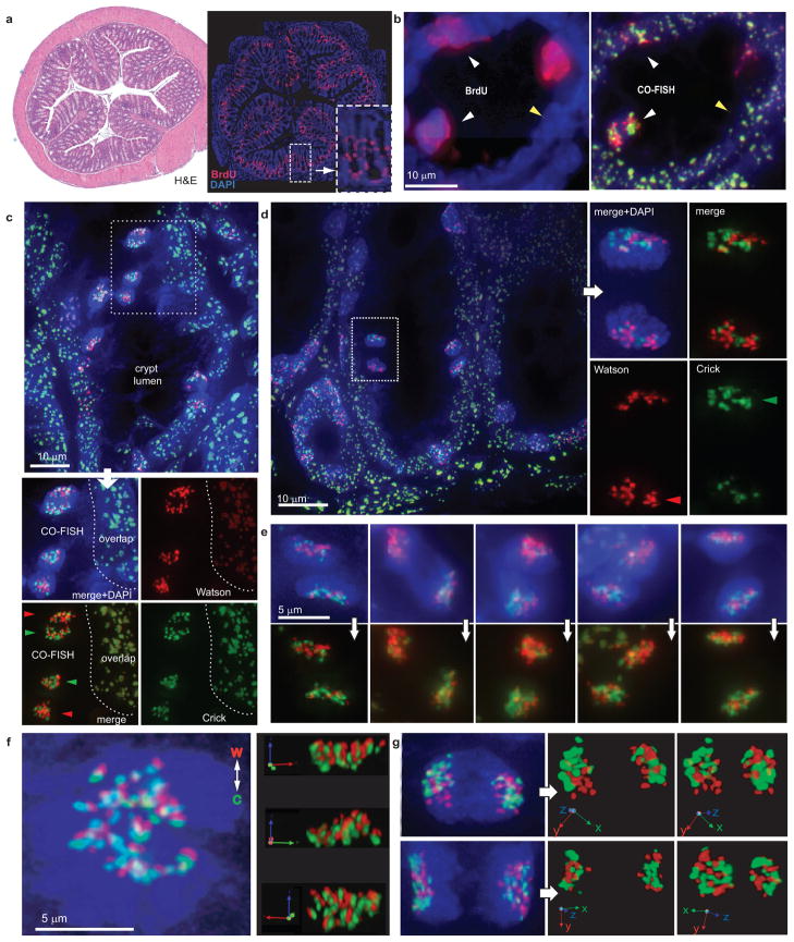
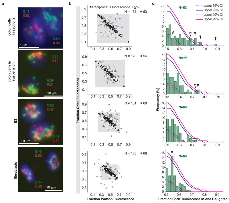
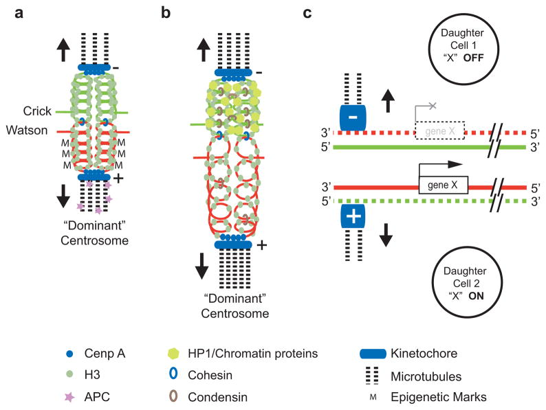
It is generally assumed that sister chromatids are genetically and functionally identical and that segregation to daughter cells is a random process. However, functional differences between sister chromatids regulate daughter cell fate in yeast and sister chromatid segregation is not random in Escherichia coli. Differentiated sister chromatids, coupled with non-random segregation, have been proposed to regulate cell fate during the development of multicellular organisms. This hypothesis has not been tested because molecular features to reliably distinguish between sister chromatids are not obvious. Here we show that parental 'Watson' and 'Crick' DNA template strands can be identified in sister chromatids of murine metaphase chromosomes using CO-FISH (chromosome orientation fluorescence in situ hybridization) with unidirectional probes specific for centromeric and telomeric repeats. All chromosomes were found to have a uniform orientation with the 5' end of the short arm on the same strand as T-rich major satellite repeats. The invariable orientation of repetitive DNA was used to differentially label sister chromatids and directly study mitotic segregation patterns in different cell types. Whereas sister chromatids appeared to be randomly distributed between daughter cells in cultured lung fibroblasts and embryonic stem cells, significant non-random sister chromatid segregation was observed in a subset of colon crypt epithelial cells, including cells outside positions reported for colon stem cells. Our results establish that DNA template sequences can be used to distinguish sister chromatids and follow their mitotic segregation in vivo.


Conflict of interest statement

The authors declare no competing financial interests.

Figures

Figure 1
Figure 1
Highly-conserved orientation of telomeric and major satellite DNA in murine chromosomes revealed by four-color CO-FISH. a, Schematic diagram of the CO-FISH procedure. b, Pseudo-color CO-FISH image of murine metaphase chromosomes. Note that major satellite repeats on all chromosomes except Y (arrow, no major satellite DNA) have the same orientation. c, Magnification of the boxed chromosome shown in b. d, Definition of “Watson” and “Crick” DNA template strands based on the uniform orientation of major satellite DNA. e, The relative distribution of Watson and Crick major satellite fluorescence can be used to study sister chromatid segregation patterns in vivo.
Figure 2
Figure 2
CO-FISH to study sister chromatid segregation patterns. a, Low magnification of adjacent colon sections stained with H&E (left panel) and DAPI and anti-BrdU antibody (right panel). b, High magnification of a section stained for BrdU (left panel) that was subsequently subjected to CO-FISH (right panel). BrdU-labeled cells show non-overlapping red and green fluorescence (white arrowheads), non-mitotic cells without BrdU show overlapping probe signals (yellow arrowhead). c, Example of CO-FISH (non-overlapping) signals in pairs of post-mitotic cells in colon crypts. d, Post-mitotic cell pairs relatively high in colon crypt with asymmetric CO-FISH fluorescence. e, Examples of asymmetric CO-FISH fluorescence in paired colon cells. f, Non-random alignment of sister chromatids at metaphase (right panels: different projections from Supplementary Movie 2). g, Mirror-image symmetry and clustered CO-FISH fluorescence in paired daughter (see also Supplementary Movies 3 and 4).
Figure 3
Figure 3
Measurements of Watson and Crick DNA template strand fluorescence in post-mitotic cells. a, Examples of fluorescence measured in the indicated cell types. b, For N cell pairs, the ratio of Watson and Crick fluorescence in one of the daughter cells (arbitrary selection) is plotted. Solid black squares show cells with reciprocal Watson and Crick fluorescence ratios ± 5%, whereas open circles represent cells with Watson/Crick fluorescence ratios outside this arbitrary cutoff. c, The observed Crick fluorescence distributions in selected individual cells (N, black squares in b) was compared to fluorescence distribution values obtained by simulated random segregation. The observed frequency (Y-axis) of Crick fluorescence (X-axis, green histograms) in one daughter cell (with the brightest Crick fluorescence) is plotted. Upper and lower 95% and 99% confidence intervals (CI, solid and dashed blue and magenta lines, respectively) represent the range of fluorescence distributions expected by chance. The values measured in colon tissues sections as well as colon cell suspensions fall outside the range for simulated random segregation (p<0.05: open arrows; p<0.01: solid arrows).
Figure 4
Figure 4
Models for the mechanism and function of asymmetric sister chromatid segregation. Only the template strand of double stranded DNA in sister chromatids is shown. a, Uneven distribution of epigenetic marks (M) between sister chromatid centromeres could result in asymmetric nucleation of microtubules or selective capture of microtubules coming from the “dominant” centrosome,. b, Differences in higher-order chromatin structure could alter the elastic properties of (peri-)centric chromatin and select specific sister chromatids via microtubules originating from the “dominant” centrosome. c, Regulation of cell fate via selective segregation of sister chromatids that differ in epigenetic marks at centromeres and selected genes.

References

    1. Klar AJ. Differentiated parental DNA strands confer developmental asymmetry on daughter cells in fission yeast. Nature. 1987;326 (6112):466–470. - PubMed
    1. White MA, Eykelenboom JK, Lopez-Vernaza MA, Wilson E, Leach DR. Non-random segregation of sister chromosomes in Escherichia coli. Nature. 2008;455 (7217):1248–1250. - PubMed
    1. Lansdorp PM. Immortal strands? Give me a break. Cell. 2007;129 (7):1244–1247. - PubMed
    1. Meyne J, Goodwin EH. Strand-specific fluorescence in situ hybridization for determining orientation and direction of DNA sequences. Methods Mol Biol. 1994;33:141–145. - PubMed
    1. Barker N, et al. Identification of stem cells in small intestine and colon by marker gene Lgr5. Nature. 2007;449 (7165):1003–1007. - PubMed

Publication types

MeSH terms