. 2009 Nov 16:1:8.
doi: 10.1186/2040-2384-1-8.

Novel insights into the differential functions of Notch ligands in vascular formation

Affiliations

Novel insights into the differential functions of Notch ligands in vascular formation

Tsutomu Kume. J Angiogenes Res. .

Abstract

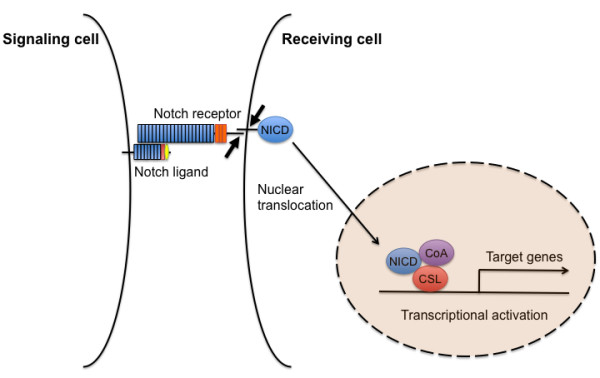
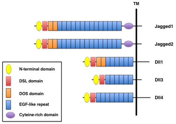
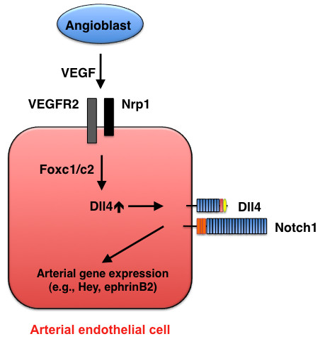
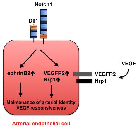
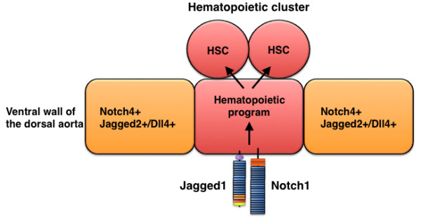
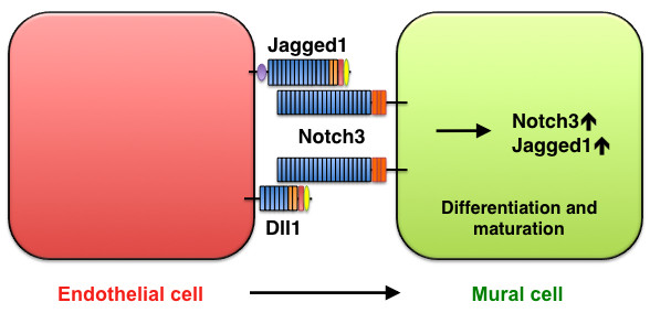
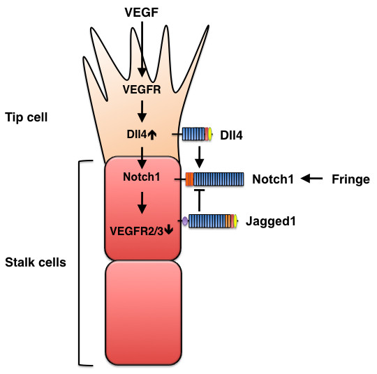
The Notch signaling pathway is a critical component of vascular formation and morphogenesis in both development and disease. Compelling evidence indicates that Notch signaling is required for the induction of arterial-cell fate during development and for the selection of endothelial tip and stalk cells during sprouting angiogenesis. In mammals, two of the four Notch receptors (Notch1 and Notch4) and three of the five Notch ligands (Jagged1, Dll1, and Dll4) are predominantly expressed in vascular endothelial cells and are important for many aspects of vascular biology. During arterial cell-fate selection and angiogenesis, the roles of Notch1 and Notch4 are thought to be similar, and the function of Dll4 is well-characterized. However, the molecular mechanisms that determine the functional similarities and differences of Notch ligands in vascular endothelial cells remain largely unknown; consequently, additional research is needed to elucidate the ligand-specific functions and mechanisms associated with Notch activation in the vascular endothelium. Results from recent studies indicate that Dll1 and Dll4 have distinct roles in the specification and maintenance of arterial cell identity, while Dll4 and Jagged1 have opposing functions in tip- and stalk-cell selection during sprouting angiogenesis. This review will focus on the newly discovered, distinct functions of several Notch ligands in the regulation of blood vessel formation and will provide perspectives for future research in the field.


Figures

Figure 1
Figure 1
A diagram of the canonical Notch signaling pathway. This schematic shows a simplified overview of the main components of Notch signaling. Upon Notch ligand binding, a two-step proteolysis cleavage process (small arrows) within the juxtamembrane region and transmembrane domain of the Notch receptor is catalyzed by a member of the disintegrin and metalloproteases (ADAMS) family and the γ-secretase containing complex, respectively, then the Notch intracellular domain (NICD) is released from the membrane and translocates to the nucleus, where it forms a transcriptional activation complex with CSL and coactivators (CoA), thereby inducing the transcription of target genes.
Figure 2
Figure 2
Domain organization of mammalian Notch ligands. Five mammalian ligands are classified into two categories, Delta-like (Dll1, Dll3, Dll4) and Serrate-like (Jagged1, Jagged2), based on structural homology to the two Drosophila ligands, Delta and Serrate. All Notch ligands have an N-terminal domain, a DSL (Delta/Serrate/LAG-2) domain and EGF-like repeats. Jagged1 and Jagged2 contain a cysteine-rich domain, whereas Jagged1, Jagged2, and Dll1 have two DOS (Delta and OSM-11-like proteins) domains located immediately following the DSL domain.
Figure 3
Figure 3
Arterial cell specification mediated by Dll4-Notch signaling. During early development, VEGF (in concert with Foxc1/c2 transcription factors) induces Dll4 expression in endothelial cells, and Dll4-Notch signaling promotes arterial gene expression.
Figure 4
Figure 4
Maintenance of arterial identity mediated by Dll1-Notch signaling. At a later stage of development, Dll1 expression is induced in arterial endothelial cells and is required for maintenance of the arterial phenotype. Dll1 also acts upstream of VEGF by regulating the expression of VEGFR2 and its co-receptor neuropilin 1 (Nrp1).
Figure 5
Figure 5
Jagged1-mediated hematopoietic program in the dorsal aorta during development. Hematopoietic stem cells (HSC) descend from Jagged1+ Notch1+ endothelial cells in the aorta.
Figure 6
Figure 6
Smooth-muscle maturation mediated by Jagged1/Dll1-Notch3 signaling. Jagged1 and Dll1 in endothelial cells activate Notch3 on mural cells, thereby promoting mural-cell maturation.
Figure 7
Figure 7
Opposing effects of Dll4 and Jagged1 on sprouting angiogenesis. VEGF signaling induces Dll4 expression in tip cells, and Dll4, in turn, activates Notch signaling in stalk cells, which reduces stalk-cell sensitivity to VEGF stimulation and, consequently suppresses the tip-cell phenotype. Conversely, Jagged1 antagonizes Dll4-mediated Notch activation in stalk cells to increase tip cell numbers and enhances vessel sprouting. The antagonistic effects of the two ligands are controlled by Fringe-dependent modulation of Notch signaling.

Similar articles

Cited by

References

    1. Bray SJ. Notch signalling: a simple pathway becomes complex. Nat Rev Mol Cell Biol. 2006;7:678–689. doi: 10.1038/nrm2009. - DOI - PubMed
    1. Alva JA, Iruela-Arispe ML. Notch signaling in vascular morphogenesis. Curr Opin Hematol. 2004;11:278–283. doi: 10.1097/01.moh.0000130309.44976.ad. - DOI - PubMed
    1. Gridley T. Notch signaling in vascular development and physiology. Development. 2007;134:2709–2718. doi: 10.1242/dev.004184. - DOI - PubMed
    1. Hofmann JJ, Iruela-Arispe ML. Notch signaling in blood vessels: who is talking to whom about what? Circ Res. 2007;100:1556–1568. doi: 10.1161/01.RES.0000266408.42939.e4. - DOI - PubMed
    1. Phng LK, Gerhardt H. Angiogenesis: a team effort coordinated by notch. Dev Cell. 2009;16:196–208. doi: 10.1016/j.devcel.2009.01.015. - DOI - PubMed

LinkOut - more resources