. 2009 Dec 16;4(12):e8328.
doi: 10.1371/journal.pone.0008328.

Phosphorylation-mediated control of histone chaperone ASF1 levels by Tousled-like kinases

Affiliations

Phosphorylation-mediated control of histone chaperone ASF1 levels by Tousled-like kinases

Maxim Pilyugin et al. PLoS One. .

Abstract

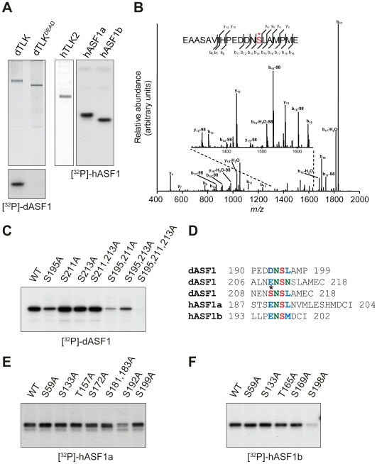
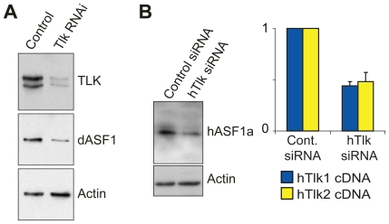
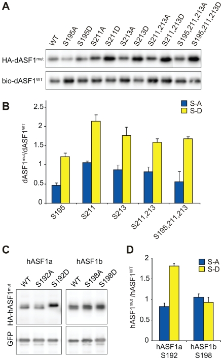
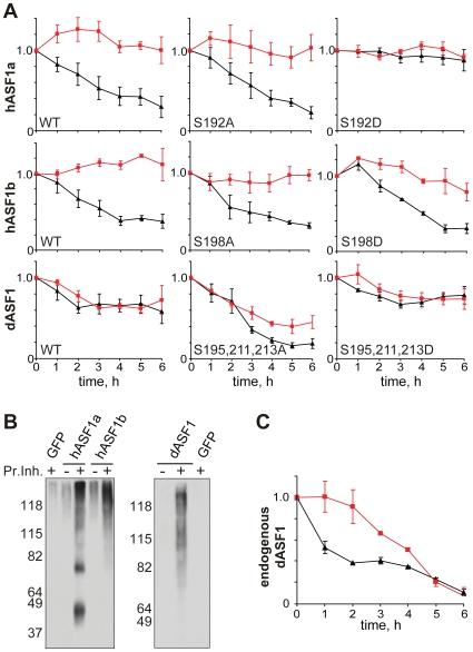
Histone chaperones are at the hub of a diverse interaction networks integrating a plethora of chromatin modifying activities. Histone H3/H4 chaperone ASF1 is a target for cell-cycle regulated Tousled-like kinases (TLKs) and both proteins cooperate during chromatin replication. However, the precise role of post-translational modification of ASF1 remained unclear. Here, we identify the TLK phosphorylation sites for both Drosophila and human ASF1 proteins. Loss of TLK-mediated phosphorylation triggers hASF1a and dASF1 degradation by proteasome-dependent and independent mechanisms respectively. Consistent with this notion, introduction of phosphorylation-mimicking mutants inhibits hASF1a and dASF1 degradation. Human hASF1b is also targeted for proteasome-dependent degradation, but its stability is not affected by phosphorylation indicating that other mechanisms are likely to be involved in control of hASF1b levels. Together, these results suggest that ASF1 cellular levels are tightly controlled by distinct pathways and provide a molecular mechanism for post-translational regulation of dASF1 and hASF1a by TLK kinases.


Conflict of interest statement

Competing Interests: The authors have declared that no competing interests exist.

Figures

Figure 1
Figure 1. Mapping of ASF1 phosphorylation sites.
(A) Drosophila TLK, TLKdead and human hTLK2 kinases were expressed in Sf9 cells and purified using His-tag. Phosphorylation of GST-ASF1 proteins by TLKs was performed in the presence of [γ-32P]ATP and revealed by autoradiography. dASF1 is phosphorylated by TLK, but not TLKdead (lower panel), and hASF1a and hASF1b can be phosphorylated by hTLK2 (right panel). (B) CID fragmentation spectrum of a doubly charged proteolytic peptide of dASF1 (m/z = 1103.96; Mascot identification peptide score  = 88). The peptide carries a phosphorylated serine residue, which is marked with an asterisk. (C) In vitro phosphorylation of dASF1 mutants by dTLK. S195A alone strongly reduces phosphorylation of dASF1, and phosphorylation of triple mutant S195/211/213A is completely abolished. (D) Alignment of ASF1 phosphorylation sites. Asterisk indicates phosphorylated S211 of dASF1 as it structurally resembles Asp (D) or Glu (E) creating optimal target site at S213 for TLK phosphorylation. (E, F) In vitro phosphorylation of hASF1a and hASF1b mutants by hTLK2. hASF1a S192A (E) and hASF1b S198A (F) mutations strongly reduce phosphorylation efficiency by hTLK2.
Figure 2
Figure 2. TLK activity is required for ASF1 stability in vivo.
(A) Depletion of dTLK from Drosophila S2 cells leads to decreased dASF1 levels. S2 cells were either mock treated or incubated with dsRNA directed against dTLK. Whole-cell extracts were prepared and analyzed by Western blotting with indicated antibodies. Actin serves as a loading control. (B) siRNA for hTLK1 and hTLK2 affects hASF1a stability in HeLa cells. Whole-cell extracts from control or siRNA treated cells were analyzed by Western blotting with anti-hASF1a and anti-Actin antibodies. Efficiency of siRNA of hTLK1 (blue) and hTLK2 (yellow) was confirmed by RT-qPCR normalized to control siRNA (right panel).
Figure 3
Figure 3. Mutations in TLK phosphorylation sites affect ASF1 protein levels.
(A) Mutated dASF1 proteins fused to HA-tag were co-expressed with wild-type bio-tagged dASF1 in S2 cells and revealed by immunoblotting. Mutated Serines within dASF1 protein are indicated. (B) Levels of mutant HA-dASF1 proteins were quantified and normalized to bio-dASF1wt protein level and compared to the same ratio obtained for HA-dASF1wt. The graph shows the mean for three experiments and error bars show standard errors of the mean (SEM). Mutated Serines are indicated (S-A mutations – blue bars, S-D – yellow bars). (C, D) Mutated HA-hASF1 proteins were co-expressed with GFP in HEK293T cells. Representative immunoblots are shown (C) and HA-hASF1 protein levels were analyzed as above (D) using GFP levels as a reference. The graph shows the mean for three experiments and error bars show SEM. Mutated Serines are indicated (S-A mutations – blue bars, S-D – yellow bars).
Figure 4
Figure 4. Phosphorylation of ASF1 hampers proteasome-dependent degradation.
(A) hASF1 and dASF1 wild type or mutant proteins were expressed in HEK293T and S2 cells respectively. Protein synthesis was blocked by cycloheximide in cells incubated with (red curves) or without (black curves) proteasome inhibitors lactacistin and MG132. ASF1 protein levels were quantified as in Figure 3 by immunoblotting for hASF1 (upper panels) and dASF1 (lower panel) every hour from 0 to 6 h and normalized to actin. All experiments were repeated at least three times and error bars show SEM. (B) HA-hASF1 and HA-dASF1 proteins or GFP were expressed in HEK293T cells. Cells were either treated (+Pr.Inh.) or not (-Pr.Inh.) with proteasome inhibitors lactacistine and MG132. Whole cell extracts were incubated with anti-HA beads and pulled-down proteins were analyzed on a western blot with anti-ubiquitin antibody. High-molecular weight smear represents poly-ubiquitinated ASF1. (C) Degradation of endogenous dASF1 dependents only partially on proteasome pathway. Protein synthesis was blocked by cycloheximide in S2 cells incubated with (red curves) or without (black curves) proteasome inhibitors lactacistin and MG132. Samples were collected every hour from 0 to 6 h. dASF1 protein was immunoblotted and quantified as above. Data are represented as mean of three experiments +/− SEM.

References

    1. De Koning L, Corpet A, Haber JE, Almouzni G. Histone chaperones: an escort network regulating histone traffic. Nat Struct Mol Biol. 2007;14:997–1007. - PubMed
    1. Eitoku M, Sato L, Senda T, Horikoshi M. Histone chaperones: 30 years from isolation to elucidation of the mechanisms of nucleosome assembly and disassembly. Cell Mol Life Sci. 2008;65:414–444. - PMC - PubMed
    1. Park YJ, Luger K. Histone chaperones in nucleosome eviction and histone exchange. Curr Opin Struct Biol. 2008;18:282–289. - PMC - PubMed
    1. Moshkin YM, Kan TW, Goodfellow H, Bezstarosti K, Maeda RK, et al. Histone chaperones ASF1 and NAP1 differentially modulate removal of active histone marks by LID-RPD3 complexes during NOTCH silencing. Mol Cell. 2009;35:782–793. - PubMed
    1. Tyler JK, Adams CR, Chen SR, Kobayashi R, Kamakaka RT, et al. The RCAF complex mediates chromatin assembly during DNA replication and repair. Nature. 1999;402:555–560. - PubMed

Publication types