. 2009 Dec 15;4(12):e8314.
doi: 10.1371/journal.pone.0008314.

Neural stem cells as a novel platform for tumor-specific delivery of therapeutic antibodies

Affiliations

Neural stem cells as a novel platform for tumor-specific delivery of therapeutic antibodies

Richard T Frank et al. PLoS One. .

Abstract

Background: Recombinant monoclonal antibodies have emerged as important tools for cancer therapy. Despite the promise shown by antibody-based therapies, the large molecular size of antibodies limits their ability to efficiently penetrate solid tumors and precludes efficient crossing of the blood-brain-barrier into the central nervous system (CNS). Consequently, poorly vascularized solid tumors and CNS metastases cannot be effectively treated by intravenously-injected antibodies. The inherent tumor-tropic properties of human neural stem cells (NSCs) can potentially be harnessed to overcome these obstacles and significantly improve cancer immunotherapy. Intravenously-delivered NSCs preferentially migrate to primary and metastatic tumor sites within and outside the CNS. Therefore, we hypothesized that NSCs could serve as an ideal cellular delivery platform for targeting antibodies to malignant tumors.

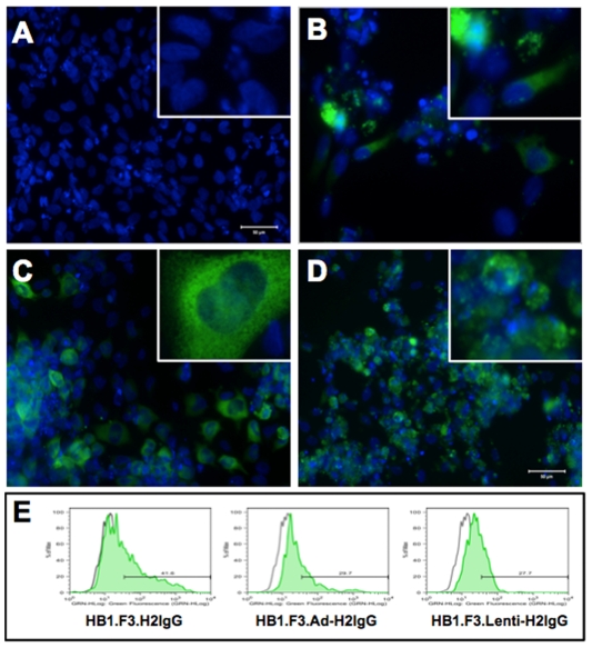
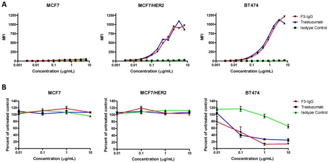
Methods and findings: As proof-of-concept, we selected Herceptin (trastuzumab), a monoclonal antibody widely used to treat HER2-overexpressing breast cancer. HER2 overexpression in breast cancer is highly correlated with CNS metastases, which are inaccessible to trastuzumab therapy. Therefore, NSC-mediated delivery of trastuzumab may improve its therapeutic efficacy. Here we report, for the first time, that human NSCs can be genetically modified to secrete anti-HER2 immunoglobulin molecules. These NSC-secreted antibodies assemble properly, possess tumor cell-binding affinity and specificity, and can effectively inhibit the proliferation of HER2-overexpressing breast cancer cells in vitro. We also demonstrate that immunoglobulin-secreting NSCs exhibit preferential tropism to tumor cells in vivo, and can deliver antibodies to human breast cancer xenografts in mice.

Conclusions: Taken together, these results suggest that NSCs modified to secrete HER2-targeting antibodies constitute a promising novel platform for targeted cancer immunotherapy. Specifically, this NSC-mediated antibody delivery system has the potential to significantly improve clinical outcome for patients with HER2-overexpressing breast cancer.


Conflict of interest statement

Competing Interests: The authors have declared that no competing interests exist.

Figures

Figure 1
Figure 1. Expression of human IgG in NSCs.
Fluorescence micrograph of parental HB1.F3 NSCs (A), HB1.F3.H2IgG (B), HB1.F3.Adeno-H2IgG NSCs (C), and HB1.F3.Lenti-H2IgG (D). Cells were stained with anti-human IgG (green) and DAPI nuclear stain (blue). Expression was confirmed by intracellular flow cytometry of fixed and permeabilized NSCs (E). Fluorescence of fixed and permeabilized parental NSCs (indicated by gray histogram) was used to set the marker for each graph.
Figure 2
Figure 2. NSC-secreted human IgG specifically binds HER2.
Transfected NSCs were co-cultured with CM-DiI-labeled (red) MCF7 (A) or MCF7/HER2 cells (B) and stained with FITC-conjugated anti-human IgG (green) and DAPI (blue). Arrows indicate NSCs expressing human IgG that does not bind to adjacent MCF7 control cells (A) or areas of NSC-secreted human IgG bound to MCF7/HER2 target cells (B). Bar, 50 µm. Flow cytometric analysis of BT474, MCF7/HER2, or MCF7 target cells after incubation with supernatant from HB1.F3.H2IgG, HB1.F3.Adeno-H2IgG, or HB1.F3.Lenti-H2IgG (C), followed by incubation with FITC-conjugated anti-human IgG (blue histograms). Red histograms on each graph show target cells incubated with supernatant from unmodified HB1.F3 NSCs as a negative control.
Figure 3
Figure 3. NSC-secreted anti-HER2 antibody is functionally equivalent to trastuzumab.
Flow cytometric analysis (A) of MCF7, MCF7/HER2, and BT474 cells labeled with F3-IgG, trastuzumab, or a human IgG isotype control antibody. Graphs show mean fluorescence intensity (MFI) of labeled cells. Inhibition of cell proliferation (B) of MCF7, MCF7/HER2, or BT474 cells treated for 6 days with F3-IgG, trastuzumab, or isotype control antibody. Graphs show proliferation as a percentage of untreated cells.
Figure 4
Figure 4. In vitro migration of NSCs to breast carcinoma conditioned media.
Migration of parental NSCs and anti-HER2-transfected HB1.F3 NSCs to breast tumor-conditioned media in an in vitro chemotaxis assay. In this assay, bovine serum albumin (BSA) was used as a negative control for chemotaxis. Both parental and transfected NSC lines preferentially migrated to MCF7/HER2 compared to negative control (2% BSA) (p<0.01).
Figure 5
Figure 5. NSCs target breast carcinoma and can deliver anti-HER2 antibody in vivo.
Confocal fluorescence micrographs of tumor sections from MCF7/HER2 xenografts. First three panels in the upper row show the presence of CM-DiI-labeled red NSCs (HB1.F3, HB1.F3.Ad-H2IgG, HB1.F3.Lenti-H2IgG, respectively) in tumors 4 days after intravenous injection. The fourth panel of the upper row shows tumor with no red NSCs in mice treated with trastuzumab alone (sporadic small red dots not associated with cells are visible as autofluorescence background). Middle row shows tumor sections stained with FITC-conjugated anti-human IgG (green). Bar, 50 µm. Insets are 2× magnification (Bar, 20 µm). Bottom row shows confirmation of the presence or absence of HB1.F3 NSCs within tumors by PCR detection of a DNA amplicon (293 bp) of the v-myc transgene, a unique identifier of the HB1.F3 cell line.

Similar articles

Cited by

References

    1. Adams GP, Schier R, McCall AM, Simmons HH, Horak EM, et al. High affinity restricts the localization and tumor penetration of single-chain fv antibody molecules. Cancer Res. 2001;61:4750–4755. - PubMed
    1. Aboody KS, Bush RA, Garcia E, Metz MZ, Najbauer J, et al. Development of a tumor-selective approach to treat metastatic cancer. PLoS ONE. 2006;1:e23. - PMC - PubMed
    1. Aboody KS, Najbauer J, Danks MK. Stem and progenitor cell-mediated tumor selective gene therapy. Gene Ther. 2008;15:739–752. - PubMed
    1. Aboody KS, Najbauer J, Schmidt NO, Yang W, Wu JK, et al. Targeting of melanoma brain metastases using engineered neural stem/progenitor cells. Neuro-oncol. 2006;8:119–126. - PMC - PubMed
    1. Brown AB, Yang W, Schmidt NO, Carroll R, Leishear KK, et al. Intravascular delivery of neural stem cell lines to target intracranial and extracranial tumors of neural and non-neural origin. Hum Gene Ther. 2003;14:1777–1785. - PubMed

Publication types

MeSH terms