. 2009 Dec 16;4(12):e8292.
doi: 10.1371/journal.pone.0008292.

Cardiac glycosides induce cell death in human cells by inhibiting general protein synthesis

Affiliations

Cardiac glycosides induce cell death in human cells by inhibiting general protein synthesis

Andrea Perne et al. PLoS One. .

Abstract

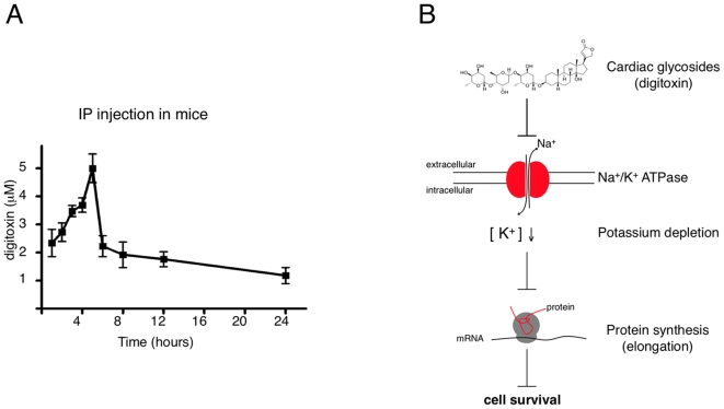
Background: Cardiac glycosides are Na(+)/K(+)-pump inhibitors widely used to treat heart failure. They are also highly cytotoxic, and studies have suggested specific anti-tumor activity leading to current clinical trials in cancer patients. However, a definitive demonstration of this putative anti-cancer activity and the underlying molecular mechanism has remained elusive.

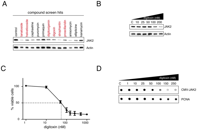
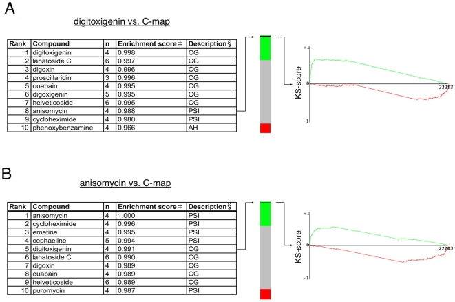
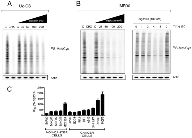
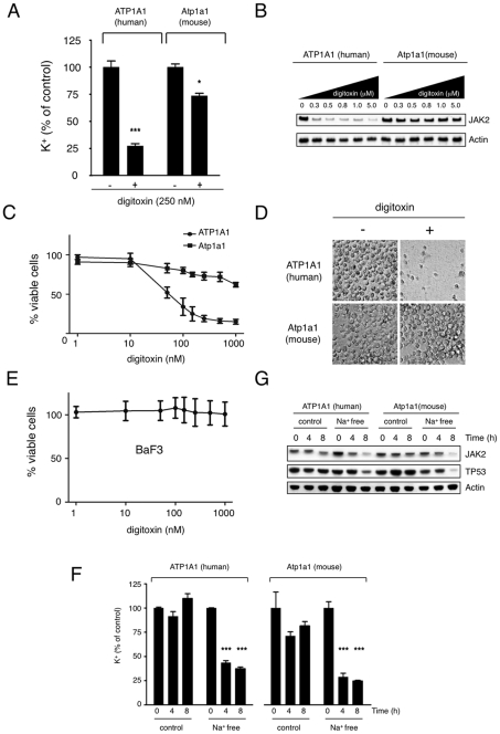
Methodology/principal findings: Using an unbiased transcriptomics approach, we found that cardiac glycosides inhibit general protein synthesis. Protein synthesis inhibition and cytotoxicity were not specific for cancer cells as they were observed in both primary and cancer cell lines. These effects were dependent on the Na(+)/K(+)-pump as they were rescued by expression of a cardiac glycoside-resistant Na(+)/K(+)-pump. Unlike human cells, rodent cells are largely resistant to cardiac glycosides in vitro and mice were found to tolerate extremely high levels.

Conclusions/significance: The physiological difference between human and mouse explains the previously observed sensitivity of human cancer cells in mouse xenograft experiments. Thus, published mouse xenograft models used to support anti-tumor activity for these drugs require reevaluation. Our finding that cardiac glycosides inhibit protein synthesis provides a mechanism for the cytotoxicity of CGs and raises concerns about ongoing clinical trials to test CGs as anti-cancer agents in humans.


Conflict of interest statement

Competing Interests: The authors have declared that no competing interests exist.

Figures

Figure 1
Figure 1. Cardiac glycosides reduce JAK2 protein levels and inhibit proliferation of JAK2 V617F mutant expressing HEL cells.
(A) Effects of compounds identified in a drug screen (1140 drugs from the Prestwick Library) on JAK2 protein expression. Human erythroleukemic HEL cells were exposed to the indicated compounds at a concentration of 5 µM for 6 hours. Cells were harvested and lysates were subjected to Western blotting using antibodies against JAK2 or actin. Compounds of the cardiac glycoside family are indicated in red. (B) HEL cells were treated with various concentrations of digitoxin as indicated for 16 hours. Equal cell numbers were harvested and expression of JAK2 and actin was determined by Western blotting. (C) HEL cells were cultured in the presence of the indicated concentrations of digitoxin (1 nM–1 µM) for 48 hours. Proliferation of cells was measured using the CellTiter-Glo assay. Results represent the mean ± S.D. of three independent experiments. (D) Human embryonic kidney cells (HEK293T) were transfected with JAK2 V617F and treated with various concentrations of digitoxin for 16 hours as indicated. Equal numbers of cells were harvested and protein expression levels of JAK2 and PCNA were quantified by dot blotting.
Figure 2
Figure 2. C-map query reveals functional similarity between CGs and protein synthesis inhibitors.
(A) A gene-expression signature for digitoxigenin was used to search for compounds with a similar mechanism of action. The ten compounds showing the strongest positive connectivity with the digitoxigenin signature from the 1,309 compounds in the C-map database, including the query compound are listed (left panel). The number of independent experiments (i.e. treatments) with each compound (n) and their set-wise enrichment scores are shown. All experiments in the C-map are rank-ordered according to connectivity score of the query signature in the “green-grey-red” barview (middle panel). All four experiments with anisomycin (black lines) cluster at the top of the green area in the bar, indicating strong and reproducible correlation with digitoxigenin. The best matching anisomycin gene-expression profile (instance id 1304) was directly compared with the gene-expression signature of digitoxigenin treated cells (right panel). The gene probes (22,283) are ordered according to differential expression of the anisomycin vs. vehicle control (left, upregulated; right, downregulated). That many of the genes regulated by anisomycin are similarly affected by digitoxigenin is illustrated by the Kolmogorov-Smirnov (KS) plot. The score (y-axis) is high at the left side of the graph because many of the genes that are upregulated (green line) by anisomycin are also upregulated by digitoxigenin. The converse is true for the downregulated genes (red line). § CG = cardiac glycoside, PSI = protein synthesis inhibitor, AH = anti-hypertensive, ± All enrichment scores have permutation p-values of <0.000001. (B) A similar query as in Figure 2a is shown but using an anisomycin signature. The best matching digitoxigenin signature (instance id 1339) was used for the Kolmogorov-Smirnov (KS) plot (right panel).
Figure 3
Figure 3. Digitoxin inhibits protein synthesis.
(A) Effects of digitoxin on 35S-Met/Cys protein incorporation in human U2-OS cells. Cells were treated with various concentrations of digitoxin for 6 hours and equal numbers of cells were subjected to 35S-Met/Cys incorporation assay as described in Materials and Methods. Actin was used as an additional loading control. (B) Dose- and time-dependent inhibition of 35S-Met/Cys uptake by digitoxin in IMR90 cells. (C) Digitoxin concentrations resulting in 50% reduction in cell number (IC50) with 95% confidence intervals (CI) obtained with neoplastic cell lines as well as with primary fibroblasts (IMR90 cells), primary human mononuclear cells (MNC, donor #1-#3) and non-tumorigenic MCF10A cells. IC50 values were calculated in GraphPad Prism 5.0 using the nonlinear regression model for dose-response (least square fitting, variable slope) and represent the results of three independent experiments in each case.
Figure 4
Figure 4. Digitoxin-induced inhibition of protein synthesis is dependent on the Na+/K+ ATPase.
(A) Effect of digitoxin on intracellular potassium levels in HEL cells with overexpression of the human or murine alpha1-chain. HEL cells transduced with ATP1A1 or Atp1a1 were incubated with digitoxin (250 nM) for 8 hours. Cells were lysed and potassium concentrations were determined (*** indicates p<0.001, * indicates p<0.05). (B) HEL cells expressing the human or murine alpha1-subunit were treated with various concentrations of digitoxin as indicated for 6 hours and the expression of JAK2 and actin was determined by Western blotting. Expression of the murine alpha-chain of the Na+K+-pump conferred resistance against the inhibitory effect of digitoxin. (C) Effects of digitoxin (1 nM–1 µM, 48 hours) on growth of ATP1A1- or Atp1a1-overexpressing HEL cells. (D) Microscope photograph of a cell culture dish containing ATP1A1- or Atp1a1-overexpressing cells after 5 days of exposure to 1 µM digitoxin. (E) Growth of murine hematopoietic Ba/F3 cells is not affected by digitoxin as assessed by CellTiter-Glo assay (three independent experiments, mean ± S.D.). (F) Effects of a sodium-free buffer on intracellular potassium levels in HEL cells overexpressing human ATP1A1 or murine Atp1a1. Cells were cultured in a sodium-free buffer or in a control buffer for up to 8 hours as indicated and potassium concentrations were measured in cell lysates. Exposure to the sodium-free buffer resulted in inhibition of the Na+/K+-pump and depletion of intracellular potassium in both cell lines (*** indicates p<0.001). (G) Inhibition of expression of JAK2 and TP53 in HEL cells exposed to sodium-free buffer. HEL cells overexpressing human ATP1A1 or murine Atp1a1 were cultured in sodium-free or control buffer for 8 hours and expression of JAK2 and TP53 was determined by Western blotting. Equal numbers of cells were loaded and actin served as an additional loading control.
Figure 5
Figure 5. Differential sensitivity of human and murine cells against CGs.
(A) Time course of digitoxin serum levels in mice injected with digitoxin (1 mg/kg) intraperitoneally. Each time point represents measurements from 5 animals (mean ± S.D.). (B) Model for the principal mechanism of action underlying the growth-inhibitory effects of cardiac glycosides. Inhibition of the Na+/K+ pump by cardiac glycosides leads to a depletion of intracellular potassium which is required for the elongation step in protein synthesis .

References

    1. Therien AG, Blostein R. Mechanisms of sodium pump regulation. Am J Physiol Cell Physiol. 2000;279:C541–566. - PubMed
    1. Prassas I, Diamandis EP. Novel therapeutic applications of cardiac glycosides. Nat Rev Drug Discov. 2008;7:926–935. - PubMed
    1. Mijatovic T, Van Quaquebeke E, Delest B, Debeir O, Darro F, et al. Cardiotonic steroids on the road to anti-cancer therapy. Biochim Biophys Acta. 2007;1776:32–57. - PubMed
    1. Lefranc F, Kiss R. The sodium pump alpha1 subunit as a potential target to combat apoptosis-resistant glioblastomas. Neoplasia. 2008;10:198–206. - PMC - PubMed
    1. Zavareh RB, Lau KS, Hurren R, Datti A, Ashline DJ, et al. Inhibition of the sodium/potassium ATPase impairs N-glycan expression and function. Cancer Res. 2008;68:6688–6697. - PubMed

Publication types