. 2010 Feb;63(2):312-21.
doi: 10.1002/mrm.22233.

3D compressed sensing for highly accelerated hyperpolarized (13)C MRSI with in vivo applications to transgenic mouse models of cancer

Affiliations

3D compressed sensing for highly accelerated hyperpolarized (13)C MRSI with in vivo applications to transgenic mouse models of cancer

Simon Hu et al. Magn Reson Med. 2010 Feb.

Abstract

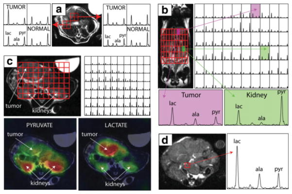
High polarization of nuclear spins in liquid state through hyperpolarized technology utilizing dynamic nuclear polarization has enabled the direct monitoring of (13)C metabolites in vivo at a high signal-to-noise ratio. Acquisition time limitations due to T(1) decay of the hyperpolarized signal require accelerated imaging methods, such as compressed sensing, for optimal speed and spatial coverage. In this paper, the design and testing of a new echo-planar (13)C three-dimensional magnetic resonance spectroscopic imaging (MRSI) compressed sensing sequence is presented. The sequence provides up to a factor of 7.53 in acceleration with minimal reconstruction artifacts. The key to the design is employing x and y gradient blips during a fly-back readout to pseudorandomly undersample k(f)-k(x)-k(y) space. The design was validated in simulations and phantom experiments where the limits of undersampling and the effects of noise on the compressed sensing nonlinear reconstruction were tested. Finally, this new pulse sequence was applied in vivo in preclinical studies involving transgenic prostate cancer and transgenic liver cancer murine models to obtain much higher spatial and temporal resolution than possible with conventional echo-planar spectroscopic imaging methods.


Figures

FIG. 1
FIG. 1
a: Simulated data set created by thresholding the peaks from a 13C phantom 3D-MRSI acquisition to remove the noise and then giving the peaks a realistic line width. The data set contained three regions, each with different chemical species. b: The simulated data set was randomly undersampled with different undersampling factors, and ℓ1 reconstructions were computed. The RMS errors between ℓ1 reconstructed data and unaccelerated data were very low for a wide range of accelerations. c: Comparison of peaks from selected voxels. Zero-fill (linear) reconstructions exhibited incoherent aliasing that had a noiselike appearance and increased with higher undersampling. ℓ1 nonlinear reconstructions with accelerations as high as 8 were nearly perfect. [Color figure can be viewed in the online issue, which is available at www.interscience.wiley.com.]
FIG. 2
FIG. 2
a: Reproduction of the noiseless data set from Fig. 1a. The three regions containing (1) alanine, (2) pyruvate/pyruvate-hydrate, and (3) lactate, respectively, are highlighted. On the right side, in the first column, those three sets are shown again but with additive white gaussian noise added. In the second column, an ℓ1 reconstruction with factor of 4 undersampling of the noisy data is shown. The third column shows the magnitude difference. b: The noise simulation in part (a) was run 100 times, with distinct noise patterns each time. For each of the peaks across the many voxels, and for both the noise added (pre-ℓ1) and noise added with factor of 4 undersampling with ℓ1 reconstruction (post-ℓ1) data sets, the mean/SD of the ratio of peak height to true noiseless peak height was plotted. All pre-ℓ1 ratios were centered on unity regardless of signal strength, but the post-ℓ1 ratios were slightly skewed downward for peaks that had low starting SNR. The bottom part shows a scatterplot of pre-ℓ1 and post-ℓ1 peak height to true noiseless peak height ratios for a selected peak over the 100 runs. [Color figure can be viewed in the online issue, which is available at www.interscience.wiley.com.]
FIG. 3
FIG. 3
a: Compressed sensing 3D-MRSI pulse sequence. Phase encode localization occurred in x/y with fly-back readout in z/f. Full echo data were collected by using twin adiabatic refocusing pulses. The key design trick was placing x/y gradient blips during the rewind portions of the fly-back readout. The blip areas were integer multiples of phase-encode steps, allowing for hopping around and random undersampling of kf-kx-ky space. b: Illustration of random undersampling of a swath of kf-kx-ky space using blips. The blips portioned out the acquisition over several lines in k-space during one pulse repetition time. c: Depiction of variable density sampling resulting in a ×7.53 accelerated sequence. Central regions of k-space were fully sampled, middle regions undersampled by a factor of 8, and outer edges undersampled by a factor of 16. d: Resulting random undersampling pattern in kf-kx-ky. [Color figure can be viewed in the online issue, which is available at www.interscience.wiley.com.]
FIG. 4
FIG. 4
Results from phantom experiments. The upper left shows an image of a slice from a cylindrical phantom with spheres containing 13C-labeled alanine, pyruvate/pyruvate-hydrate, and lactate. The adjacent boxes show comparisons of spectral grids from 3D-MRSI acquisitions with no undersampling, ×3.37 undersampling, and ×7.53 undersampling. The 16 × 16 voxels shown have 3.75 mm × 3.75 mm in-plane resolution. The 6 × 6 grids to the right show spectra from the alanine sphere only (frequency axis zoomed in). The ℓ1 reconstructions for the accelerated acquisitions matched the unaccelerated acquisition, showing high spectral quality and the preservation of small peaks. [Color figure can be viewed in the online issue, which is available at www.interscience.wiley.com.]
FIG. 5
FIG. 5
a: In vivo validation in a transgenic mouse model of prostate cancer showing a comparison of spectra from an unaccelerated acquisition and an accelerated one with a quarter the voxel size and acquired in about the same time. The accelerated acquisition with higher resolution allowed for better delineation of the mouse body and better depiction of tumor heterogeneity. The spectra in the accelerated acquisition were of high quality, and small peaks were preserved. The color overlay maps generated from the accelerated spectra show high-intensity regions as brightly colored and highlight the spatial localization of metabolites according to tissue type. Tumor and nontumor regions showed clear differences in metabolic profile, and the boundaries were clearly delineated. b: A comparison of spectra from a different slice in the same mouse, with the higher-resolution spectra depicting heterogeneity and the boundaries of a metastasis better than the lower-resolution spectra. The full 3D acquisition allowed for imaging disease in multiple slices. c: High-quality spectra from a different prostate cancer mouse in which the ×7.53 accelerated sequence was used to quadruple resolution and nearly halve acquisition time. Once again, small peaks were preserved. [Color figure can be viewed in the online issue, which is available at www.interscience.wiley.com.]
FIG. 6
FIG. 6
a: Spectra from a transgenic liver cancer mouse with an early-stage tumor, as shown in the upper left of the anatomic image, in which acceleration was used to reduce voxel size by a factor of 4 (×3.37 acceleration, 0.034-cm3 voxel size, 16-sec acquisition). Tumor voxels exhibited dramatically elevated lactate/pyruvate ratios. The higher resolution reduced partial voluming such that distinct metabolic profiles were observed in tumor and adjacent tissue voxels. b: A separate data set in the same mouse with the same acquisition parameters in which the 3D-MRSI data are presented coronally to emphasize the distinct metabolic profiles in tumor and other tissues. c: A 3D-MRSI data set from a different mouse with a moderate-stage tumor at the level of the kidneys. Distinct differences between tumor and normal tissue are readily visualized in the color overlay maps. d: Data from a mouse with a large very late-stage tumor. Elevated alanine, as well as lactate, was detected in the tumor mass.

References

    1. Ardenkjaer-Larsen JH, Fridlund B, Gram A, Hansson G, Hansson L, Lerche MH, Servin R, Thaning M, Golman K. Increase in signal-to-noise ratio of >10,000 times in liquid-state NMR. Proc Natl Acad Sci USA. 2003;100:10158–10163. - PMC - PubMed
    1. Wolber J, Ellner F, Fridlund B, Gram A, Johannesson H, Hansson G, Hansson LH, Lerche MH, Mansson S, Servin R, Thaning M, Golman K, Ardenkjaer-Larsen JH. Generating highly polarized nuclear spins in solution using dynamic nuclear polarization. Nucl Instrum Methods Phys Res A. 2004;526:173–181.
    1. Golman K, Zandt R, Thaning M. Real-time metabolic imaging. Proc Natl Acad Sci USA. 2006;103:11270–11275. - PMC - PubMed
    1. Golman K, Peterson JS. Metabolic imaging and other applications of hyperpolarized 13C. Acad Radiol. 2006;13:932–942. - PubMed
    1. Golman K, Zandt RI, Lerche M, Pehrson R, Ardenkjaer-Larsen JH. Metabolic imaging by hyperpolarized 13C magnetic resonance imaging for in vivo tumor diagnosis. Cancer Res. 2006;66:10855–10860. - PubMed

Publication types