

A stimulatory thyrotropin receptor antibody (M22) and thyrotropin increase interleukin-6 expression and secretion in Graves' orbital preadipocyte fibroblasts
- PMID: 20017620
- PMCID: PMC2833174
- DOI: 10.1089/thy.2009.0278
A stimulatory thyrotropin receptor antibody (M22) and thyrotropin increase interleukin-6 expression and secretion in Graves' orbital preadipocyte fibroblasts
Abstract
Background: Patients with Graves' ophthalmopathy (GO) have circulating autoantibodies directed against the thyrotropin receptor (TSHR) and elevated levels of the proinflammatory cytokine interleukin-6 (IL-6) in both serum and orbital tissues. We hypothesized that these autoantibodies might increase IL-6 expression and secretion in preadipocyte fibroblasts and adipocytes from patients with GO, and thus directly impact the clinical activity of the disease.
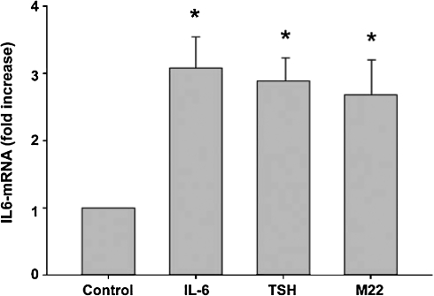
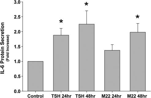
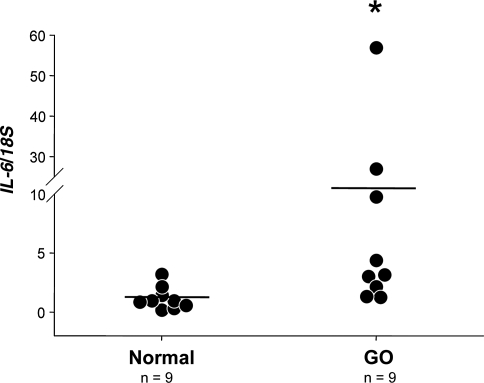
Methods: IL-6 mRNA levels were measured in cultures of GO orbital preadipocytes (n = 3) treated during adipocyte differentiation with a monoclonal stimulatory TSHR antibody (M22; 10 ng/mL), IL-6 (1 ng/mL), or TSH (10 U/L). Additionally, levels of IL-6 protein secretion were assessed after adipocyte differentiation in orbital cultures exposed to TSH or M22 for 24 or 48 hours (n = 8). IL-6 mRNA levels were also measured in orbital adipose tissue specimens from well-characterized GO patients (n = 9) and normal individuals (n = 9).
Results: Treatment of GO orbital preadipocyte cultures with IL-6, TSH, or M22 during adipocyte differentiation resulted in increased IL-6 mRNA levels (3.1-fold, 2.9-fold, and 2.7-fold, respectively; p < 0.05). Treatment of orbital cultures with M22 or TSH after adipocyte differentiation enhanced the release of IL-6 protein into the medium at both 24 and 48 hours for TSH (mean 1.9- and 2.3-fold; p = 0.002 and 0.015, respectively) and at 48 hours for M22 (mean 2.0-fold; p = 0.005). In addition, we found mean IL-6 mRNA levels to be significantly increased in GO orbital adipose tissue specimens (10-fold; p < 0.01), primarily attributable to high levels in three of the four patients with clinical activity scores >or=5.
Conclusions: Both TSH and M22 increase IL-6 expression in orbital preadipocyte fibroblasts and IL-6 secretion by mature adipocytes. These results suggest that circulating TSHR autoantibodies in GO might play a direct role in the clinical activity of the disease.
Figures
Similar articles
-
A stimulatory TSH receptor antibody enhances adipogenesis via phosphoinositide 3-kinase activation in orbital preadipocytes from patients with Graves' ophthalmopathy.J Mol Endocrinol. 2011 Apr 12;46(3):155-63. doi: 10.1530/JME-11-0006. Print 2011 Jun. J Mol Endocrinol. 2011. PMID: 21321093 Free PMC article.
-
Forkhead Transcription Factor FOXO1 Is Regulated by Both a Stimulatory Thyrotropin Receptor Antibody and Insulin-Like Growth Factor-1 in Orbital Fibroblasts from Patients with Graves' Ophthalmopathy.Thyroid. 2015 Oct;25(10):1145-50. doi: 10.1089/thy.2015.0254. Epub 2015 Aug 19. Thyroid. 2015. PMID: 26213859 Free PMC article.
-
Interleukin-6 stimulates thyrotropin receptor expression in human orbital preadipocyte fibroblasts from patients with Graves' ophthalmopathy.Thyroid. 2001 Oct;11(10):929-34. doi: 10.1089/105072501753210984. Thyroid. 2001. PMID: 11716039
-
Current perspectives on the role of orbital fibroblasts in the pathogenesis of Graves' ophthalmopathy.Exp Eye Res. 2016 Jan;142:83-91. doi: 10.1016/j.exer.2015.02.007. Exp Eye Res. 2016. PMID: 26675405 Review.
-
Thyrotropin receptor expression in orbital adipose/connective tissues from patients with thyroid-associated ophthalmopathy.Thyroid. 2002 Mar;12(3):193-5. doi: 10.1089/105072502753600124. Thyroid. 2002. PMID: 11952038 Review.
Cited by
-
Steroid-Resistant Graves' Orbitopathy Treated with Tocilizumab in Real-World Clinical Practice: A 9-Year Single-Center Experience.J Clin Med. 2021 Feb 11;10(4):706. doi: 10.3390/jcm10040706. J Clin Med. 2021. PMID: 33670151 Free PMC article.
-
A review of TSHR- and IGF-1R-related pathogenesis and treatment of Graves' orbitopathy.Front Immunol. 2023 Jan 19;14:1062045. doi: 10.3389/fimmu.2023.1062045. eCollection 2023. Front Immunol. 2023. PMID: 36742308 Free PMC article. Review.
-
Immunopathogenesis of Graves' ophthalmopathy: the role of the TSH receptor.Best Pract Res Clin Endocrinol Metab. 2012 Jun;26(3):281-9. doi: 10.1016/j.beem.2011.10.003. Best Pract Res Clin Endocrinol Metab. 2012. PMID: 22632365 Free PMC article. Review.
-
Differential profiling of lacrimal cytokines in patients suffering from thyroid-associated orbitopathy.Sci Rep. 2018 Jul 17;8(1):10792. doi: 10.1038/s41598-018-29113-2. Sci Rep. 2018. PMID: 30018377 Free PMC article.
-
Allosteric modulators hit the TSH receptor.Endocrinology. 2014 Jan;155(1):1-5. doi: 10.1210/en.2013-2079. Endocrinology. 2014. PMID: 24364583 Free PMC article. No abstract available.
References
-
- Heufelder AE. Dutton CM. Sarkar G. Donovan KA. Bahn RS. Detection of TSH receptor RNA in cultured fibroblasts from patients with Graves' ophthalmopathy and pretibial dermopathy. Thyroid. 1993;3:297–300. - PubMed
-
- Feliciello A. Porcellini A. Ciullo I. Bonavolonta G. Avvedimento EV. Fenzi G. Expression of thyrotropin-receptor mRNA in healthy and Graves' disease retro-orbital tissue. Lancet. 1993;342:337–338. - PubMed
-
- Ludgate M. Crisp M. Lane C. Costagliola S. Vassart G. Weetman A. Daunerie C. Many MC. The thyrotropin receptor in thyroid eye disease. Thyroid. 1998;8:411–413. - PubMed
-
- Mengistu M. Lukes YG. Nagy EV. Burch HB. Carr FE. Lahiri S. Burman KD. TSH receptor gene expression in retroocular fibroblasts. J Endocrinol Invest. 1994;17:437–441. - PubMed
-
- Bahn RS. Dutton CM. Natt N. Joba W. Spitzweg C. Heufelder AE. Thyrotropin receptor expression in Graves' orbital adipose/connective tissues: potential autoantigen in Graves' ophthalmopathy. J Clin Endocrinol Metab. 1998;83:998–1002. - PubMed
Publication types
MeSH terms
Substances
Grants and funding
LinkOut - more resources
Full Text Sources
