. 2010 Jan 15;201(2):190-8.
doi: 10.1086/649228.

Enhanced memory responses to seasonal H1N1 influenza vaccination of the skin with the use of vaccine-coated microneedles

Affiliations

Enhanced memory responses to seasonal H1N1 influenza vaccination of the skin with the use of vaccine-coated microneedles

Yeu-Chun Kim et al. J Infect Dis. .

Abstract

Background: Morbidity and mortality due to influenza could be reduced by improved vaccination.

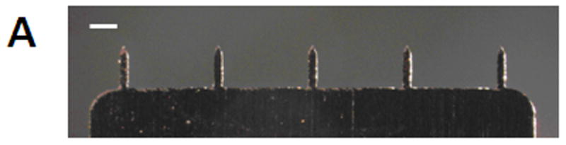
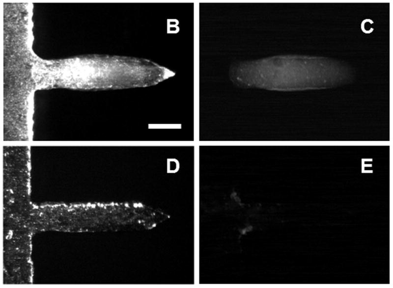
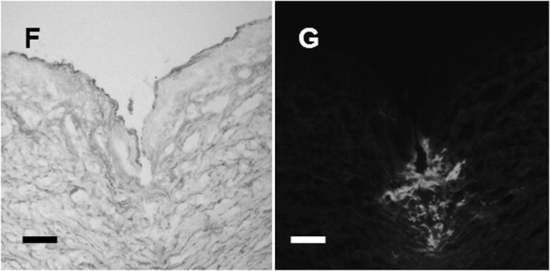
Methods: To develop a novel skin delivery method that is simple and allows for easy self-administration, we prepared microneedle patches with stabilized influenza vaccine and investigated their protective immune responses.

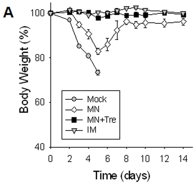
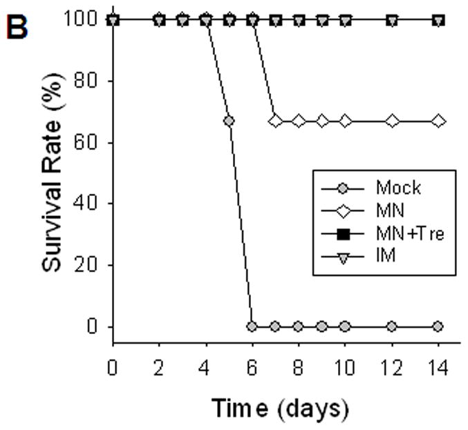
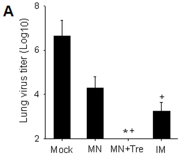
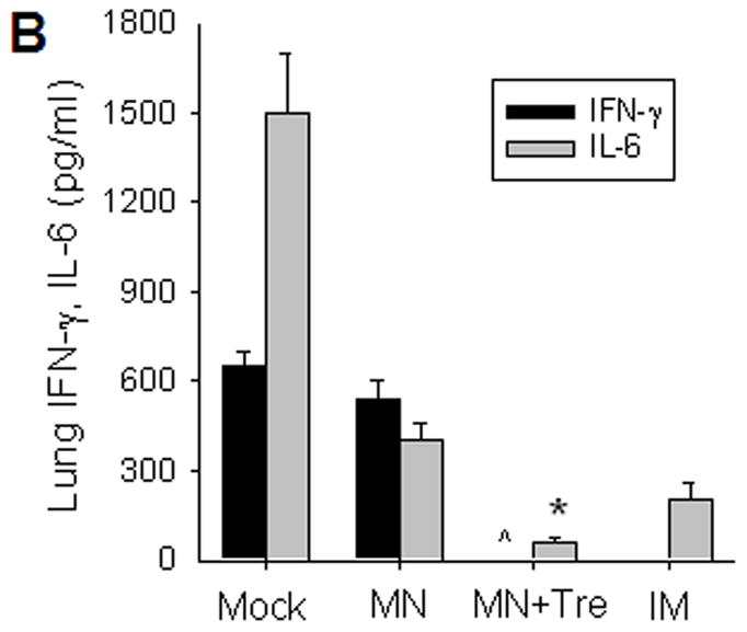
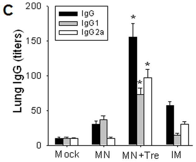
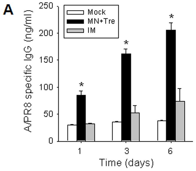
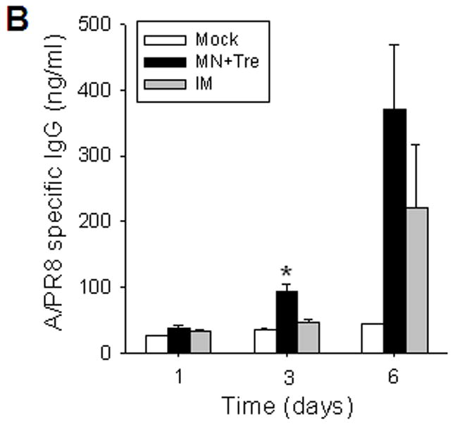
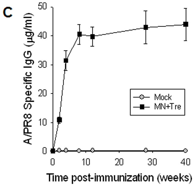
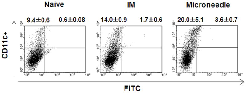
Results: Mice vaccinated with a single microneedle dose of trehalose-stabilized influenza vaccine developed strong antibody responses that were long-lived. Compared with traditional intramuscular vaccination, stabilized microneedle vaccination was superior in inducing protective immunity, as was evidenced by efficient clearance of virus from the lung and enhanced humoral and antibody-secreting cell immune responses after 100% survival from lethal challenge. Vaccine stabilization was found to be important, because mice vaccinated with an unstabilized microneedle vaccine elicited a weaker immunoglobulin G 2a antibody response, compared with the stabilized microneedle vaccine, and were only partially protected against viral challenge. Improved trafficking of dendritic cells to regional lymph nodes as a result of microneedle delivery to the skin might play a role in contributing to improved protective immunity.

Conclusions: These findings suggest that vaccination of the skin using a microneedle patch can improve protective efficacy and induce long-term sustained immunogenicity and may also provide a simple method of administration to improve influenza vaccination coverage.


Conflict of interest statement

Potential conflict of interests: M.P. is an inventor on patents (one of which has been licensed) and a consultant and advisor to companies working on microneedles. Although there are currently no products based on microneedles, and this study does not involve any commercial products, the results of this research could indirectly influence the success of possible future commercial activities in which M.P. has an interest.

Figures

Figure 1
Figure 1
Microneedle coated with influenza vaccine. (A) Image of a 5-microneedle array (scale bar = 500 μm). Bright-field (B, D) and fluorescence (C, E) micrographs of a microneedle coated with red-fluorescent inactivated influenza virus before (B, C) and 10 min after (D, E) insertion into human cadaver skin (scale bar = 200 μm). Histologic section of human cadaver skin fixed after insertion of a vaccine-coated microneedle imaged by (F) bright-field microscopy showing skin deformation and needle track across epidermis and into superficial dermis and (G) fluorescence microscopy showing deposition of red-fluorescent vaccine coating in skin (scale bar = 200 μm).
Figure 1
Figure 1
Microneedle coated with influenza vaccine. (A) Image of a 5-microneedle array (scale bar = 500 μm). Bright-field (B, D) and fluorescence (C, E) micrographs of a microneedle coated with red-fluorescent inactivated influenza virus before (B, C) and 10 min after (D, E) insertion into human cadaver skin (scale bar = 200 μm). Histologic section of human cadaver skin fixed after insertion of a vaccine-coated microneedle imaged by (F) bright-field microscopy showing skin deformation and needle track across epidermis and into superficial dermis and (G) fluorescence microscopy showing deposition of red-fluorescent vaccine coating in skin (scale bar = 200 μm).
Figure 2
Figure 2
Protection against lethal challenge infection. Immunized mice were challenged with a lethal dose (20 LD50) of a highly pathogenic A/PR8 influenza virus 5 weeks after a single vaccination (n=10). (A) Body weight change. (B) Survival rates were monitored daily for 14 days (n=6). Similar survival rates were obtained in two independent experiments indicating reproducible results. Mock, microneedle immunization without vaccine; MN, microneedle immunization with influenza vaccine formulated in the absence of trehalose; MN+Tre, microneedle immunization with influenza vaccine formulated in the presence of trehalose (15%); IM, intramuscular immunization with unprocessed influenza vaccine. Dead animals were removed and only live animals were counted for the body weight analysis, reflecting the rebound in body weight as a result of recovery. For the analysis at day 4 post challenge, 4 out of 10 mice were sacrificed and the remaining 6 mice were monitored.
Figure 2
Figure 2
Protection against lethal challenge infection. Immunized mice were challenged with a lethal dose (20 LD50) of a highly pathogenic A/PR8 influenza virus 5 weeks after a single vaccination (n=10). (A) Body weight change. (B) Survival rates were monitored daily for 14 days (n=6). Similar survival rates were obtained in two independent experiments indicating reproducible results. Mock, microneedle immunization without vaccine; MN, microneedle immunization with influenza vaccine formulated in the absence of trehalose; MN+Tre, microneedle immunization with influenza vaccine formulated in the presence of trehalose (15%); IM, intramuscular immunization with unprocessed influenza vaccine. Dead animals were removed and only live animals were counted for the body weight analysis, reflecting the rebound in body weight as a result of recovery. For the analysis at day 4 post challenge, 4 out of 10 mice were sacrificed and the remaining 6 mice were monitored.
Figure 3
Figure 3
Protective efficacy of microneedle vaccination. (A) Lung virus titers. Lungs from individual mice were extracted (1 ml media per mouse lung, n=4, *p<0.05, + p<0.01). The detection limit for lung viral titers was 50 pfu per 1 ml lung extracts of individual mice. (B) Lung inflammatory IL-6 and IFN-γ cytokines (n=4, *p<0.05, ^p<0.05). (C) Virus specific antibodies in lungs (n=4, *p<0.05). Lungs were collected from individual mice at day 4 post challenge and antibody levels determined by ELISA were expressed end-point dilution titers. Groups of mice are as described in the legend of Figure 2. *: MN+Tre compared with Mock, MN, and IM. ^: MN+Tre compared with Mock, MN. +: MN+Tre compared with Mock.
Figure 3
Figure 3
Protective efficacy of microneedle vaccination. (A) Lung virus titers. Lungs from individual mice were extracted (1 ml media per mouse lung, n=4, *p<0.05, + p<0.01). The detection limit for lung viral titers was 50 pfu per 1 ml lung extracts of individual mice. (B) Lung inflammatory IL-6 and IFN-γ cytokines (n=4, *p<0.05, ^p<0.05). (C) Virus specific antibodies in lungs (n=4, *p<0.05). Lungs were collected from individual mice at day 4 post challenge and antibody levels determined by ELISA were expressed end-point dilution titers. Groups of mice are as described in the legend of Figure 2. *: MN+Tre compared with Mock, MN, and IM. ^: MN+Tre compared with Mock, MN. +: MN+Tre compared with Mock.
Figure 4
Figure 4
Rapid recall and long-term immune responses. Bone marrow and spleen cells were harvested at day 4 post challenge (n=4), and kinetics of virus-specific IgG antibody production were determined. Antibody levels in the fourfold diluted in vitro culture supernatants were determined by ELISA (OD at 450 nm) after 1 to 6 days of incubation, and were expressed in concentrations (ng/ml) using standard mouse antibodies. (A) Bone marrow cell cultures (5 × 10⁵ cells/well) in the absence of influenza virus antigen stimulation (n=4, *p<0.05). (B) Spleen cell cultures (5 × 10⁵ cells/well) in the plate coated with inactivated influenza viral antigen (n=4, *p<0.005). Groups of mice (n=4, *p<0.05) are as described in the legend of Figure 2. (C) Long-term maintenance of antibody levels by microneedle vaccination. In an independent experiment for long-term antibody responses, virus specific antibody responses were determined over a 9 month period in mice (n=6) immunized in the skin with trehalose-formulated microneedle vaccine (0.7 μg inactivated influenza virus). Time 0 is the IgG value from the serum samples obtained before immunization of mice with microneedle vaccine. Serial diluted serum samples were used for ELISA and antibody levels were expressed in concentrations (μg/ml) from a mouse antibody standard curve. *: MN+Tre compared with Mock and IM.
Figure 4
Figure 4
Rapid recall and long-term immune responses. Bone marrow and spleen cells were harvested at day 4 post challenge (n=4), and kinetics of virus-specific IgG antibody production were determined. Antibody levels in the fourfold diluted in vitro culture supernatants were determined by ELISA (OD at 450 nm) after 1 to 6 days of incubation, and were expressed in concentrations (ng/ml) using standard mouse antibodies. (A) Bone marrow cell cultures (5 × 10⁵ cells/well) in the absence of influenza virus antigen stimulation (n=4, *p<0.05). (B) Spleen cell cultures (5 × 10⁵ cells/well) in the plate coated with inactivated influenza viral antigen (n=4, *p<0.005). Groups of mice (n=4, *p<0.05) are as described in the legend of Figure 2. (C) Long-term maintenance of antibody levels by microneedle vaccination. In an independent experiment for long-term antibody responses, virus specific antibody responses were determined over a 9 month period in mice (n=6) immunized in the skin with trehalose-formulated microneedle vaccine (0.7 μg inactivated influenza virus). Time 0 is the IgG value from the serum samples obtained before immunization of mice with microneedle vaccine. Serial diluted serum samples were used for ELISA and antibody levels were expressed in concentrations (μg/ml) from a mouse antibody standard curve. *: MN+Tre compared with Mock and IM.
Figure 5
Figure 5
DC migration to the draining lymph nodes. After one day treatment of mice (n=5) with microneedle or IM delivery of FITC, the inquinal lymph nodes were harvested, and CD11c+ and CD11c+FITC+ DC populations were analyzed by flow cytometry. Percentages of gated populations in the upper quadrants of each dot plot are shown. The plots are representative from two independent experiments.

Similar articles

Cited by

References

    1. Cox RJ, Brokstad KA, Ogra P. Influenza virus: Immunity and vaccination strategies. Comparison of the immune response to inactivated and live, attenuated influenza vaccines. Scandinavian J Immunol. 2004;59:1–15. - PubMed
    1. Palese P. Influenza: Old and new threats. Nat Med. 2004;10:S82–7. - PubMed
    1. Centers for Disease Control and Prevention. Prevention and control of influenza, recommendations of the Advisory Committee on Immunization Practices. Morbidity and Mortality Weekly Report. 2008;57:1–60. - PubMed
    1. Kermode M. Unsafe injections in low-income country health settings: need for injection safety promotion to prevent the spread of blood-borne viruses. Health Promot Int. 2004;19:95–103. - PubMed
    1. Mitragotri S. Immunization without needles. Nat Rev Immunol. 2005;5:905–16. - PubMed

Publication types

Substances