. 2009;11(6):R194.
doi: 10.1186/ar2893. Epub 2009 Dec 17.

Interleukin-23 is critical for full-blown expression of a non-autoimmune destructive arthritis and regulates interleukin-17A and RORgammat in gammadelta T cells

Affiliations

Interleukin-23 is critical for full-blown expression of a non-autoimmune destructive arthritis and regulates interleukin-17A and RORgammat in gammadelta T cells

Ferry Cornelissen et al. Arthritis Res Ther. 2009.

Abstract

Introduction: Interleukin (IL)-23 is essential for the development of various experimental autoimmune models. However, the role of IL-23 in non-autoimmune experimental arthritis remains unclear. Here, we examined the role of IL-23 in the non-autoimmune antigen-induced arthritis (AIA) model. In addition, the regulatory potential of IL-23 in IL-17A and retinoic acid-related orphan receptor gamma t (RORgammat) expression in CD4+ and TCRgammadelta+ T cells was evaluated systemically as well as at the site of inflammation.

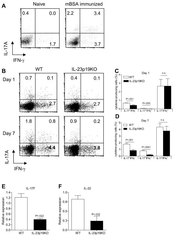
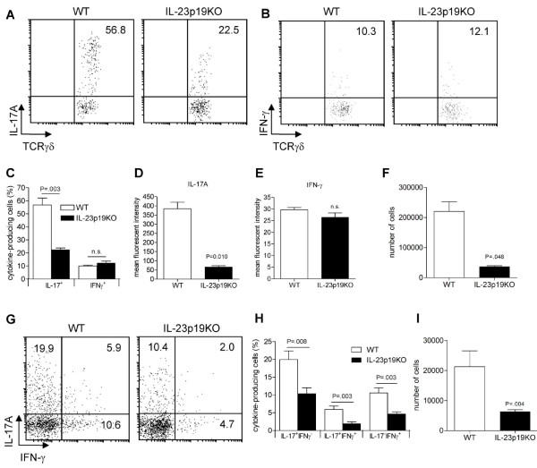
Methods: Antigen-induced arthritis was induced in wild-type, IL-23p19-deficient and IL-17 Receptor A - knockout mice. At different time points, synovial cytokine and chemokine expression was measured. At days 1 and 7 of AIA, splenocytes and joint-infiltrating cells were isolated and analyzed for intracellular IL-17A and interferon (IFN)-gamma ex-vivo by flow cytometry. In splenic CD4+ and TCRgammadelta+ T cells gene expression was quantified by flow cytometry and quantitative PCR.

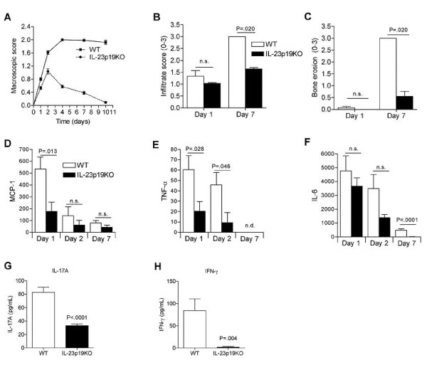
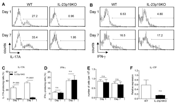
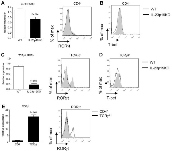
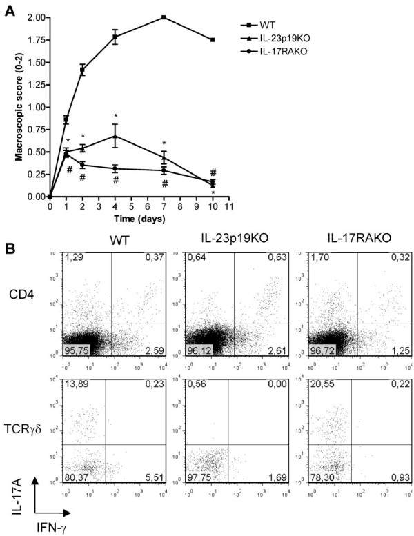
Results: IL-23 was critical for full-blown AIA. Lack of IL-23 did not prevent the onset of joint inflammation but stopped the progression to a destructive synovitis. IL-23 regulated IL-17A expression in CD4+ T cells in the spleen. Of note, IL-17A and IFN-gamma expression was reduced in CD4+ T cells in the inflamed joints of IL-23p19-deficient mice. Interestingly, IL-23 was also critical for the induction of IL-17A and RORgammat but not IFN-gamma in TCRgammadelta+ T cells in the inflamed joints. The importance of the IL-23/IL-17 axis was further confirmed using IL-17 Receptor A knockout mice showing significantly milder AIA compared to control mice, with a disease course comparable to that of IL-23p19-deficient mice.

Conclusions: These data show that IL-23 is critical for full-blown expression of a non-autoimmune destructive arthritis and regulates the proportion of IL-17A and IFN-gamma-positive CD4+ T cells at the site of inflammation. Furthermore, IL-23 regulates IL-17A and RORgammat expression in TCRgammadelta T cells in arthritis. These findings indicate that regulating the IL-23 pathway may have therapeutic potential in non-autoimmune arthritis.


Figures

Figure 1
Figure 1
IL-23 has a critical role in the progression of antigen-induced arthritis. WT and IL-23p19KO mice were immunized with mBSA/CFA and one week later mono-arthritis was induced by injecting mBSA directly into the knee joints. A. Arthritis score was determined macroscopically at different time points. Mean values and SEM are given for 8 to 31 mice per group. Data are obtained from three separate experiments. * P < 0.001, WT vs IL-23p19KO. B, Histological analyses of joint inflammation and, C, bone erosion after H&E staining. B and C, Mean values and SEM are given from two separate experiments with a total of 20 knee joints per group. D-F. Cytokine levels in synovial washouts taken at days 1, 2 and 7. G and H. IL-17A and IFN-γ levels in synovial washouts taken at day 1. Mean values and SEM are given for 5 to 10 washouts obtained from two separate experiments.
Figure 2
Figure 2
The induction of Th17 cells in AIA is IL-23 mediated. A. WT mice were immunized with mBSA/CFA and 10 days later splenocytes were isolated and stimulated for four hours with PMA/Ionomycin, gated for CD4+ T cells and analyzed for intracellular IL-17A and IFN-γ expression. Numbers indicate percentage of positive cells within each quadrant. B-D. Antigen-induced arthritis was induced in WT and IL-23p19KO mice and at days 1 and 7, after i.a. mBSA injection, the splenocytes were isolated and stimulated for 4 h with PMA/Ionomycin and analyzed for intracellular IL-17A and IFN-γ expression on a CD4+ T cell gate. B. Numbers indicate percentage of positive cells within each quadrant. C and D. Quantification of flow cytrometric analyses from B; mean values and SEM are given for 6 to 12 mice per group from two to four independent experiments. E and F. On day 7 of AIA, splenic CD3+CD4+ T cells were FACS-sorted and gene expression was analyzed by quantitative RT-PCR for IL-17F and IL-22 respectively. Mean values and SEM are given for three mice. P-values were calculated using the student t-test.
Figure 3
Figure 3
IL-23 is critical for the induction of IL-17A and RORγt in TCRγδ T cells. A and B. At days 1 and 7 of AIA, splenocytes were isolated and stimulated for four hours with PMA/Ionomycin, gated for CD3+TCRγδ+ T cells and analyzed for intracellular IL-17A and IFN-γ respectively. Numbers in quadrants indicate percentage of cytokine-positive cells. C and D. Quantification of flow cytrometric analyses from A and B for IL-17A and IFN-γ; mean values and SEM are given for three to six mice for day 1 and for nine mice for day 7. E. Cell counts of total amount of TCRγδ+ T cells present in spleen on day 1 and 7 of AIA in WT and IL-23p19KO mice. F. On day 7 of AIA, splenic CD3+TCRγδ+ T cells were FACS-sorted and IL-17F gene expression was analyzed by quantitative RT-PCR. Mean values and SEM are given for three mice. P-values were calculated using the student's t-test.
Figure 4
Figure 4
IL-23 is essential for RORγt expression in TCRγδ + T cells. At day 7 of AIA, WT and IL-23p19KO mice were sacrificed and splenocytes were isolated. A. CD3+CD4+ T cells were FACS-sorted and RORγt gene expression was analyzed by quantitative RT-PCR (left panel); RORγt was measured intracellular and CD4+ T cells were gated (right panel). B T-bet expression was measured intracellular and CD4+ T cells were gated. C. CD3+TCRγδ+ T cells were FACS-sorted and RORγt gene expression was analyzed by quantitative RT-PCR (left panel); RORγt was measured intracellular and γδ+ T cells were gated (right panel). D. T-bet expression was measured intracellular and γδ+ T cells were gated. E. Comparison of the mRNA quantification of RORγt expression between FACS-sorted CD4+ and γδ+ T cells. Mean values and SEM are given for three mice and P-values were calculated using the student's t-test.
Figure 5
Figure 5
IL-23 deficiency results in less IL-17 production in the inflamed joint. At day 7 of AIA, cells from the arthritic joints of WT and IL-23p19KO mice were isolated and stimulated for four hours with PMA/Ionomycin. A. Intracellular cytokine staining of TCRγδ+ T cells for IL-17. B. Intracellular cytokine staining of TCRγδ+ T cells for IFN-γ; data from A and B are representatives from three mice per group. C. Quantification of flow cytrometric analyses from A and B. D. MFI was calculated for TCRγδ+ IL-17+ T cells. E. MFI was calculated for TCRγδ+ IFN-γ+ T cells. F. Total numbers of TCRγδ+ T cells in the joints of arthritic WT and IL-23p19KO mice. C-F, Mean values and SEM are given for three mice and P-values were calculated using the student t-test. G. Intracellular cytokine staining of CD4+ T cells for IL-17A and IFN-γ. Data are representatives of six mice per group. H. Quantification of flow cytrometric analyses from G. I. Total numbers of CD4+ T cells present in arthritic joints from WT and IL-23p19KO mice.
Figure 6
Figure 6
IL-17 receptor signaling is critical for the progression of AIA. A. WT, IL-23p19KO and IL-17RAKO mice were immunized with mBSA/CFA and mono-arthritis was induced one week later by an i.a. injection of mBSA. At days 1, 2, 4, 7 and 10 macroscopic scores were assessed. Mean values and SEM are given for six mice per group. * P < 0.01 WT vs IL-23P19KO and #, P < 0.01 WT vs IL-17RAKO. B. At day 7 of AIA, splenocytes from WT, IL-23p19KO and IL-17RAKO were isolated and stimulated for four hours with PMA/Ionomycin and analyzed for intracellular IL-17A and IFN-γ expression on a CD4+ and TCRγδ+ T cell gate. Numbers indicate percentage of positive cells within each quadrant and each dot plot shows a representative mouse from three mice per group analyzed.

Similar articles

Cited by

References

    1. Oppmann B, Lesley R, Blom B, Timans JC, Xu Y, Hunte B, Vega F, Yu N, Wang J, Singh K, Zonin F, Vaisberg E, Churakova T, Liu M, Gorman D, Wagner J, Zurawski S, Liu Y, Abrams JS, Moore KW, Rennick D, de Waal-Malefyt R, Hannum C, Bazan JF, Kastelein RA. Novel p19 protein engages IL-12p40 to form a cytokine, IL-23, with biological activities similar as well as distinct from IL-12. Immunity. 2000;13:715–725. doi: 10.1016/S1074-7613(00)00070-4. - DOI - PubMed
    1. Hunter CA. New IL-12-family members: IL-23 and IL-27, cytokines with divergent functions. Nat Rev Immunol. 2005;5:521–531. doi: 10.1038/nri1648. - DOI - PubMed
    1. Cua DJ, Sherlock J, Chen Y, Murphy CA, Joyce B, Seymour B, Lucian L, To W, Kwan S, Churakova T, Zurawski S, Wiekowski M, Lira SA, Gorman D, Kastelein RA, Sedgwick JD. Interleukin-23 rather than interleukin-12 is the critical cytokine for autoimmune inflammation of the brain. Nature. 2003;421:744–748. doi: 10.1038/nature01355. - DOI - PubMed
    1. Murphy CA, Langrish CL, Chen Y, Blumenschein W, McClanahan T, Kastelein RA, Sedgwick JD, Cua DJ. Divergent pro- and antiinflammatory roles for IL-23 and IL-12 in joint autoimmune inflammation. J Exp Med. 2003;198:1951–1957. doi: 10.1084/jem.20030896. - DOI - PMC - PubMed
    1. Langrish CL, McKenzie BS, Wilson NJ, de Waal Malefyt R, Kastelein RA, Cua DJ. IL-12 and IL-23: master regulators of innate and adaptive immunity. Immunol Rev. 2004;202:96–105. doi: 10.1111/j.0105-2896.2004.00214.x. - DOI - PubMed

Publication types

MeSH terms

Substances