

Argon: neuroprotection in in vitro models of cerebral ischemia and traumatic brain injury
- PMID: 20017934
- PMCID: PMC2811924
- DOI: 10.1186/cc8214
Argon: neuroprotection in in vitro models of cerebral ischemia and traumatic brain injury
Abstract
Introduction: Recently, it has been shown in several experimental settings that the noble gases xenon and helium have neuroprotective properties. In this study we tested the hypothesis that the noble gas argon has a neuroprotective potential as well. Since traumatic brain injury and stroke are widespread and generate an enormous economic and social burden, we investigated the possible neuroprotective effect in in vitro models of traumatic brain injury and cerebral ischemia.
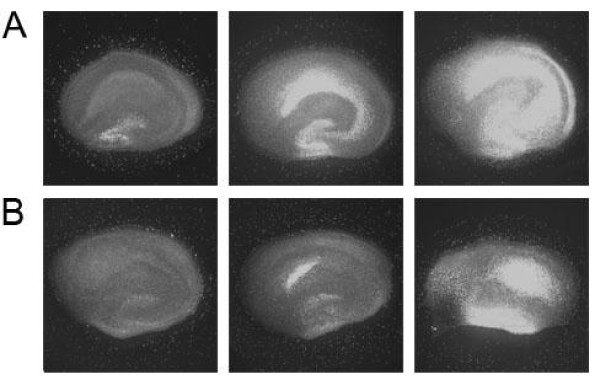
Methods: Organotypic hippocampal slice cultures from mice pups were subjected to either oxygen-glucose deprivation or to a focal mechanical trauma and subsequently treated with three different concentrations (25, 50 and 74%) of argon immediately after trauma or with a two-or-three-hour delay. After 72 hours of incubation tissue injury assessment was performed using propidium iodide, a staining agent that becomes fluorescent when it diffuses into damaged cells via disintegrated cell membranes.
Results: We could show argon's neuroprotective effects at different concentrations when applied directly after oxygen-glucose deprivation or trauma. Even three hours after application, argon was still neuroprotective.
Conclusions: Argon showed a neuroprotective effect in both in vitro models of oxygen-glucose deprivation and traumatic brain injury. Our promising results justify further in vivo animal research.
Figures


Comment in
-
Argon neuroprotection.Crit Care. 2010;14(1):117. doi: 10.1186/cc8847. Epub 2010 Feb 22. Crit Care. 2010. PMID: 20236500 Free PMC article.
Similar articles
-
Neuroprotection against traumatic brain injury by xenon, but not argon, is mediated by inhibition at the N-methyl-D-aspartate receptor glycine site.Anesthesiology. 2013 Nov;119(5):1137-48. doi: 10.1097/ALN.0b013e3182a2a265. Anesthesiology. 2013. PMID: 23867231
-
Noble gas neuroprotection: xenon and argon protect against hypoxic-ischaemic injury in rat hippocampus in vitro via distinct mechanisms.Br J Anaesth. 2019 Nov;123(5):601-609. doi: 10.1016/j.bja.2019.07.010. Epub 2019 Aug 27. Br J Anaesth. 2019. PMID: 31470983 Free PMC article.
-
Propofol: neuroprotection in an in vitro model of traumatic brain injury.Crit Care. 2009;13(2):R61. doi: 10.1186/cc7795. Epub 2009 Apr 27. Crit Care. 2009. PMID: 19397790 Free PMC article.
-
Argon: systematic review on neuro- and organoprotective properties of an "inert" gas.Int J Mol Sci. 2014 Oct 10;15(10):18175-96. doi: 10.3390/ijms151018175. Int J Mol Sci. 2014. PMID: 25310646 Free PMC article.
-
Noble gases and neuroprotection: summary of current evidence.Curr Opin Anaesthesiol. 2021 Oct 1;34(5):603-606. doi: 10.1097/ACO.0000000000001033. Curr Opin Anaesthesiol. 2021. PMID: 34224430 Review.
Cited by
-
Dexmedetomidine is neuroprotective in an in vitro model for traumatic brain injury.BMC Neurol. 2012 Apr 11;12:20. doi: 10.1186/1471-2377-12-20. BMC Neurol. 2012. PMID: 22494498 Free PMC article.
-
Application of medical gases in the field of neurobiology.Med Gas Res. 2011 Jun 27;1(1):13. doi: 10.1186/2045-9912-1-13. Med Gas Res. 2011. PMID: 22146102 Free PMC article.
-
Desflurane impairs outcome of organotypic hippocampal slices in an in vitro model of traumatic brain injury.Med Gas Res. 2016 Apr 4;6(1):3-9. doi: 10.4103/2045-9912.179338. eCollection 2016 Mar. Med Gas Res. 2016. PMID: 27826417 Free PMC article.
-
Neuroprotective properties of levosimendan in an in vitro model of traumatic brain injury.BMC Neurol. 2010 Oct 21;10:97. doi: 10.1186/1471-2377-10-97. BMC Neurol. 2010. PMID: 20964834 Free PMC article.
-
Bustling argon: biological effect.Med Gas Res. 2013 Oct 3;3(1):22. doi: 10.1186/2045-9912-3-22. Med Gas Res. 2013. PMID: 24088583 Free PMC article.
References
-
- Behnke AR, Yarbrough OD. Respiratory resistance, oil-water solubility, and mental effects of argon, compared with helium and nitrogen. Am J Physiol. 1939;126:409–415.
-
- Soldatov PE, D'Iachenko AI, Pavlov BN, Fedotov AP, Chuguev AP. [Survival of laboratory animals in argon-containing hypoxic gaseous environments] Aviakosm Ekolog Med. 1998;32:33–37. - PubMed
-
- Yarin YM, Amarjargal N, Fuchs J, Haupt H, Mazurek B, Morozova SV, Gross J. Argon protects hypoxia-, cisplatin- and gentamycin-exposed hair cells in the newborn rat's organ of Corti. Hear Res. 2005;201:1–9. - PubMed
-
- Jawad N, Rizvi M, Gu J, Adeyi O, Tao G, Maze M, Ma D. Neuroprotection (and lack of neuroprotection) afforded by a series of noble gases in an in vitro model of neuronal injury. Neurosci Lett. 2009;460:232–236. - PubMed
-
- Ma D, Wilhelm S, Maze M, Franks NP. Neuroprotective and neurotoxic properties of the 'inert' gas, xenon. Br J Anaesth. 2002;89:739–746. - PubMed
Publication types
MeSH terms
Substances
LinkOut - more resources
Full Text Sources
Other Literature Sources
