

A taxonomy of epithelial human cancer and their metastases
- PMID: 20017941
- PMCID: PMC2806369
- DOI: 10.1186/1755-8794-2-69
A taxonomy of epithelial human cancer and their metastases
Abstract
Background: Microarray technology has allowed to molecularly characterize many different cancer sites. This technology has the potential to individualize therapy and to discover new drug targets. However, due to technological differences and issues in standardized sample collection no study has evaluated the molecular profile of epithelial human cancer in a large number of samples and tissues. Additionally, it has not yet been extensively investigated whether metastases resemble their tissue of origin or tissue of destination.
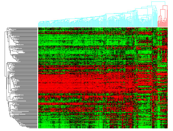
Methods: We studied the expression profiles of a series of 1566 primary and 178 metastases by unsupervised hierarchical clustering. The clustering profile was subsequently investigated and correlated with clinico-pathological data. Statistical enrichment of clinico-pathological annotations of groups of samples was investigated using Fisher exact test. Gene set enrichment analysis (GSEA) and DAVID functional enrichment analysis were used to investigate the molecular pathways. Kaplan-Meier survival analysis and log-rank tests were used to investigate prognostic significance of gene signatures.
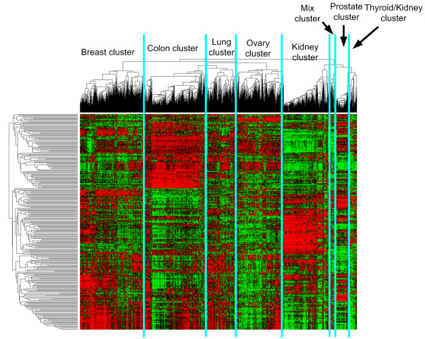
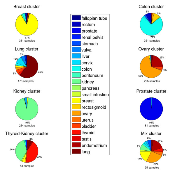
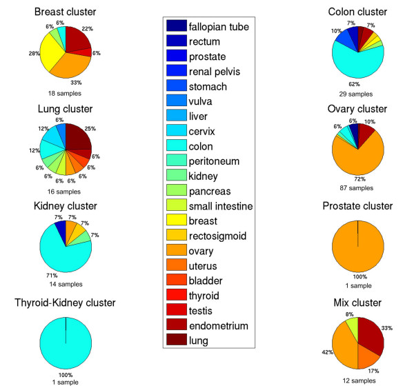
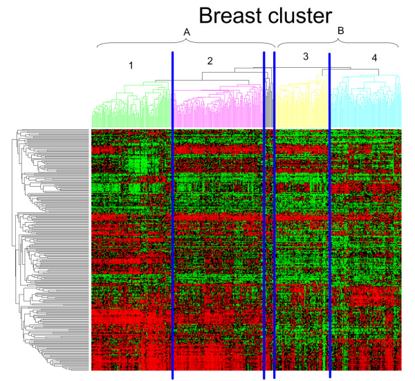
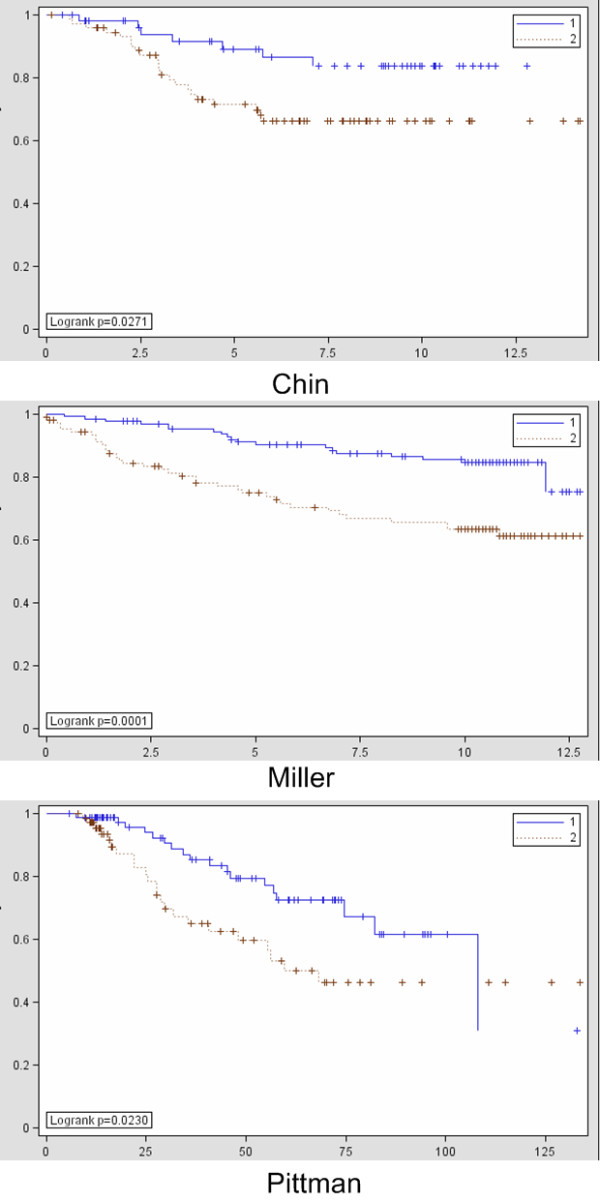
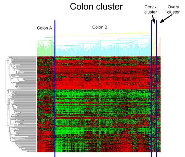
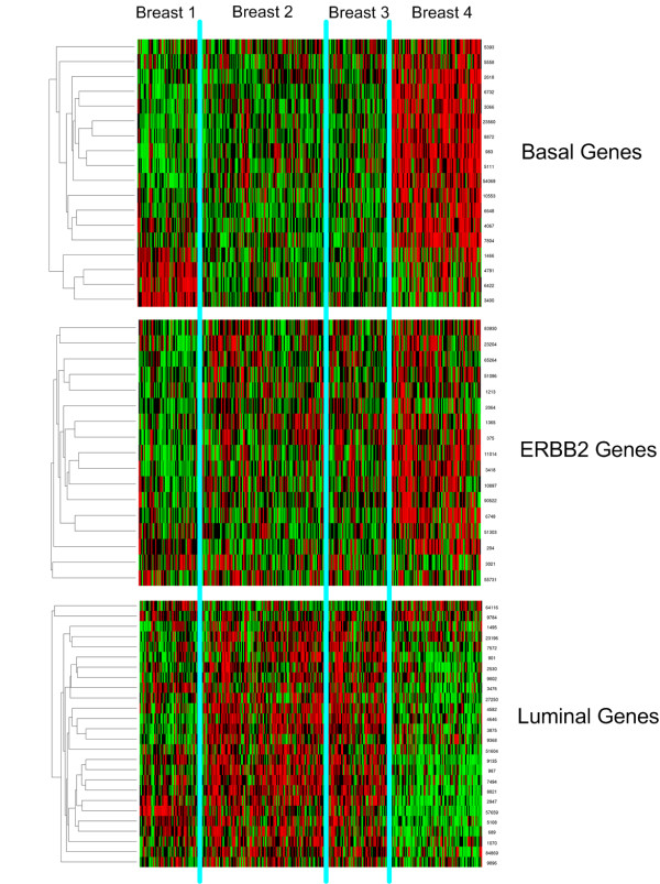
Results: Large clusters corresponding to breast, gastrointestinal, ovarian and kidney primary tissues emerged from the data. Chromophobe renal cell carcinoma clustered together with follicular differentiated thyroid carcinoma, which supports recent morphological descriptions of thyroid follicular carcinoma-like tumors in the kidney and suggests that they represent a subtype of chromophobe carcinoma. We also found an expression signature identifying primary tumors of squamous cell histology in multiple tissues. Next, a subset of ovarian tumors enriched with endometrioid histology clustered together with endometrium tumors, confirming that they share their etiopathogenesis, which strongly differs from serous ovarian tumors. In addition, the clustering of colon and breast tumors correlated with clinico-pathological characteristics. Moreover, a signature was developed based on our unsupervised clustering of breast tumors and this was predictive for disease-specific survival in three independent studies. Next, the metastases from ovarian, breast, lung and vulva cluster with their tissue of origin while metastases from colon showed a bimodal distribution. A significant part clusters with tissue of origin while the remaining tumors cluster with the tissue of destination.
Conclusion: Our molecular taxonomy of epithelial human cancer indicates surprising correlations over tissues. This may have a significant impact on the classification of many cancer sites and may guide pathologists, both in research and daily practice. Moreover, these results based on unsupervised analysis yielded a signature predictive of clinical outcome in breast cancer. Additionally, we hypothesize that metastases from gastrointestinal origin either remember their tissue of origin or adapt to the tissue of destination. More specifically, colon metastases in the liver show strong evidence for such a bimodal tissue specific profile.
Figures
Similar articles
-
FOXM1 expression is significantly associated with chemotherapy resistance and adverse prognosis in non-serous epithelial ovarian cancer patients.J Exp Clin Cancer Res. 2017 May 8;36(1):63. doi: 10.1186/s13046-017-0536-y. J Exp Clin Cancer Res. 2017. PMID: 28482906 Free PMC article.
-
[Significance and expression of PAX8, PAX2, p53 and RAS in ovary and fallopian tubes to origin of ovarian high grade serous carcinoma].Zhonghua Fu Chan Ke Za Zhi. 2017 Oct 25;52(10):687-696. doi: 10.3760/cma.j.issn.0529-567X.2017.10.008. Zhonghua Fu Chan Ke Za Zhi. 2017. PMID: 29060967 Chinese.
-
Patterns of gene expression in different histotypes of epithelial ovarian cancer correlate with those in normal fallopian tube, endometrium, and colon.Clin Cancer Res. 2005 Sep 1;11(17):6116-26. doi: 10.1158/1078-0432.CCR-04-2509. Clin Cancer Res. 2005. PMID: 16144910
-
Morphologic, Immunophenotypic, and Molecular Features of Epithelial Ovarian Cancer.Oncology (Williston Park). 2016 Feb;30(2):166-76. Oncology (Williston Park). 2016. PMID: 26892153 Review.
-
miRNA-200a/c as potential biomarker in epithelial ovarian cancer (EOC): evidence based on miRNA meta-signature and clinical investigations.Oncotarget. 2016 Dec 6;7(49):81621-81633. doi: 10.18632/oncotarget.13154. Oncotarget. 2016. PMID: 27835595 Free PMC article. Review.
Cited by
-
TP53 mutations detected in circulating tumor cells present in the blood of metastatic triple negative breast cancer patients.Breast Cancer Res. 2014 Oct 9;16(5):445. doi: 10.1186/s13058-014-0445-3. Breast Cancer Res. 2014. PMID: 25307991 Free PMC article.
-
Identifying master regulators of cancer and their downstream targets by integrating genomic and epigenomic features.Pac Symp Biocomput. 2013:123-34. Pac Symp Biocomput. 2013. PMID: 23424118 Free PMC article.
-
Gastric and duodenal squamous cell carcinoma: metastatic or primary?World J Surg Oncol. 2013 Aug 19;11(1):204. doi: 10.1186/1477-7819-11-204. World J Surg Oncol. 2013. PMID: 23957943 Free PMC article.
-
Gene signatures in hepatocellular carcinoma (HCC).Semin Cancer Biol. 2011 Feb;21(1):4-9. doi: 10.1016/j.semcancer.2010.09.002. Epub 2010 Sep 17. Semin Cancer Biol. 2011. PMID: 20851183 Free PMC article. Review.
-
High Mobility Group A proteins in esophageal carcinomas.Cell Cycle. 2016 Sep 16;15(18):2410-3. doi: 10.1080/15384101.2016.1215388. Epub 2016 Aug 2. Cell Cycle. 2016. PMID: 27484584 Free PMC article.
References
Publication types
MeSH terms
LinkOut - more resources
Full Text Sources
