

Acute dosing of latrepirdine (Dimebon), a possible Alzheimer therapeutic, elevates extracellular amyloid-beta levels in vitro and in vivo
- PMID: 20017949
- PMCID: PMC2806870
- DOI: 10.1186/1750-1326-4-51
Acute dosing of latrepirdine (Dimebon), a possible Alzheimer therapeutic, elevates extracellular amyloid-beta levels in vitro and in vivo
Abstract
Background: Recent reports suggest that latrepirdine (Dimebon, dimebolin), a retired Russian antihistamine, improves cognitive function in aged rodents and in patients with mild to moderate Alzheimer's disease (AD). However, the mechanism(s) underlying this benefit remain elusive. AD is characterized by extracellular accumulation of the amyloid-beta (Abeta) peptide in the brain, and Abeta-lowering drugs are currently among the most popular anti-amyloid agents under development for the treatment of AD. In the current study, we assessed the effect of acute dosing of latrepirdine on levels of extracellular Abeta using in vitro and in vivo experimental systems.
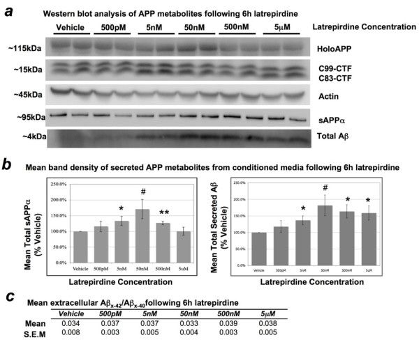
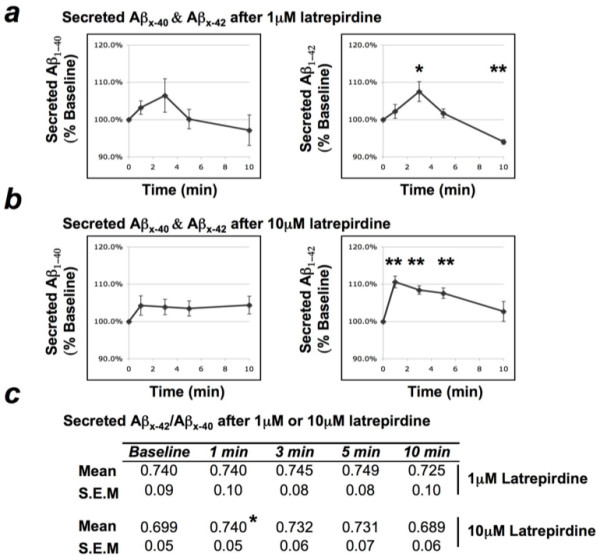
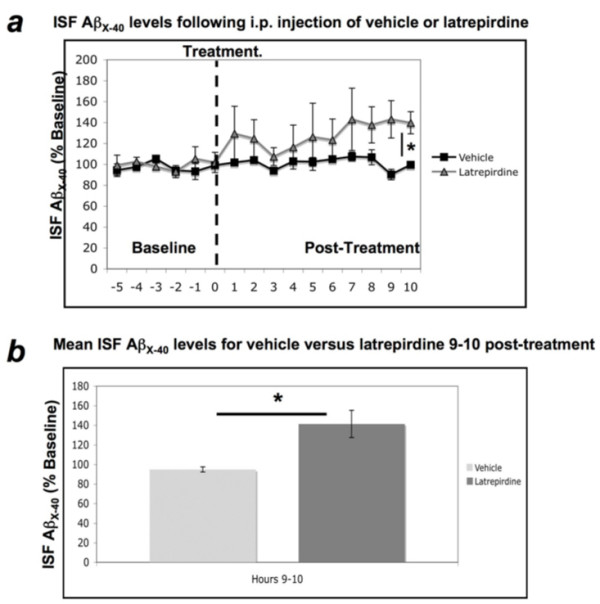
Results: We evaluated extracellular levels of Abeta in three experimental systems, under basal conditions and after treatment with latrepirdine. Mouse N2a neuroblastoma cells overexpressing Swedish APP were incubated for 6 hr in the presence of either vehicle or vehicle + latrepirdine (500pM-5 muM). Synaptoneurosomes were isolated from TgCRND8 mutant APP-overexpressing transgenic mice and incubated for 0 to 10 min in the absence or presence of latrepirdine (1 muM or 10 muM). Drug-naïve Tg2576 Swedish mutant APP overexpressing transgenic mice received a single intraperitoneal injection of either vehicle or vehicle + latrepirdine (3.5 mg/kg). Picomolar to nanomolar concentrations of acutely administered latrepirdine increased the extracellular concentration of Abeta in the conditioned media from Swedish mutant APP-overexpressing N2a cells by up to 64% (p = 0.01), while a clinically relevant acute dose of latrepirdine administered i.p. led to an increase in the interstitial fluid of freely moving APP transgenic mice by up to 40% (p = 0.01). Reconstitution of membrane protein trafficking and processing is frequently inefficient, and, consistent with this interpretation, latrepirdine treatment of isolated TgCRND8 synaptoneurosomes involved higher concentrations of drug (1-10 muM) and led to more modest increases in extracellular Abeta(x-42 )levels (+10%; p = 0.001); of note, however, was the observation that extracellular Abeta(x-40 )levels did not change.
Conclusions: Here, we report the surprising association of acute latrepirdine dosing with elevated levels of extracellular Abeta as measured in three independent neuron-related or neuron-derived systems, including the hippocampus of freely moving Tg2576 mice. Given the reported association of chronic latrepirdine treatment with improvement in cognitive function, the effects of chronic latrepirdine treatment on extracellular Abeta levels must now be determined.
Figures
Similar articles
-
Latrepirdine (Dimebon®), a potential Alzheimer therapeutic, regulates autophagy and neuropathology in an Alzheimer mouse model.Autophagy. 2013 Apr;9(4):617-8. doi: 10.4161/auto.23487. Epub 2013 Feb 4. Autophagy. 2013. PMID: 23380933 Free PMC article.
-
Dimebon alters hippocampal amyloid pathology in 3xTg-AD mice.Int J Physiol Pathophysiol Pharmacol. 2012;4(3):115-27. Epub 2012 Sep 20. Int J Physiol Pathophysiol Pharmacol. 2012. PMID: 23071869 Free PMC article.
-
Latrepirdine improves cognition and arrests progression of neuropathology in an Alzheimer's mouse model.Mol Psychiatry. 2013 Aug;18(8):889-97. doi: 10.1038/mp.2012.106. Epub 2012 Jul 31. Mol Psychiatry. 2013. PMID: 22850627 Free PMC article.
-
Recent Advances in the Modeling of Alzheimer's Disease.Front Neurosci. 2022 Mar 31;16:807473. doi: 10.3389/fnins.2022.807473. eCollection 2022. Front Neurosci. 2022. PMID: 35431779 Free PMC article. Review.
-
Cognitive Impairment in Transgenic Mouse Models of Amyloid Deposition.In: Levin ED, Buccafusco JJ, editors. Animal Models of Cognitive Impairment. Boca Raton (FL): CRC Press/Taylor & Francis; 2006. Chapter 10. In: Levin ED, Buccafusco JJ, editors. Animal Models of Cognitive Impairment. Boca Raton (FL): CRC Press/Taylor & Francis; 2006. Chapter 10. PMID: 21204370 Free Books & Documents. Review.
Cited by
-
Dimebolin in dementia.CNS Neurosci Ther. 2011 Jun;17(3):199-205. doi: 10.1111/j.1755-5949.2010.00156.x. Epub 2010 Jun 11. CNS Neurosci Ther. 2011. PMID: 20553307 Free PMC article. Review.
-
Repetitive Low-Level Blast Exposure Improves Behavioral Deficits and Chronically Lowers Aβ42 in an Alzheimer Disease Transgenic Mouse Model.J Neurotrauma. 2021 Nov 15;38(22):3146-3173. doi: 10.1089/neu.2021.0184. Epub 2021 Sep 15. J Neurotrauma. 2021. PMID: 34353119 Free PMC article.
-
Mechanism matters.Nat Med. 2010 Apr;16(4):347. doi: 10.1038/nm0410-347. Nat Med. 2010. PMID: 20376007 No abstract available.
-
Herbal/Natural Compounds Resist Hallmarks of Brain Aging: From Molecular Mechanisms to Therapeutic Strategies.Antioxidants (Basel). 2023 Apr 13;12(4):920. doi: 10.3390/antiox12040920. Antioxidants (Basel). 2023. PMID: 37107295 Free PMC article. Review.
-
Identification of the Potential Gene Regulatory Networks and Therapeutics in Aged Mice With Postoperative Neurocognitive Disorder.Front Neurosci. 2021 Jun 24;15:689188. doi: 10.3389/fnins.2021.689188. eCollection 2021. Front Neurosci. 2021. PMID: 34248489 Free PMC article.
References
-
- Shankar GM, Li S, Mehta TH, Garcia-Munoz A, Shepardson NE, Smith I, Brett FM, Farrell MA, Rowan MJ, Lemere CA, Regan CM, Walsh DM, Sabatini BL, Selkoe DJ. Amyloid-beta protein dimers isolated directly from Alzheimer's brains impair synaptic plasticity and memory. Nat Med. 2008;14(8):837–842. doi: 10.1038/nm1782. - DOI - PMC - PubMed
Grants and funding
LinkOut - more resources
Full Text Sources
Other Literature Sources
Medical
