. 2009 Dec 18:9:69.
doi: 10.1186/1471-213X-9-69.

Characterization of the stem cell system of the acoel Isodiametra pulchra

Affiliations

Characterization of the stem cell system of the acoel Isodiametra pulchra

Katrien De Mulder et al. BMC Dev Biol. .

Abstract

Background: Tissue plasticity and a substantial regeneration capacity based on stem cells are the hallmark of several invertebrate groups such as sponges, cnidarians and Platyhelminthes. Traditionally, Acoela were seen as an early branching clade within the Platyhelminthes, but became recently positioned at the base of the Bilateria. However, little is known on how the stem cell system in this new phylum is organized. In this study, we wanted to examine if Acoela possess a neoblast-like stem cell system that is responsible for development, growth, homeostasis and regeneration.

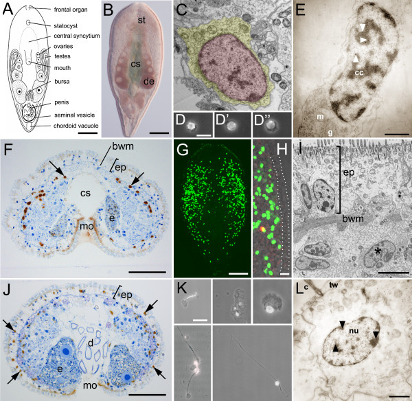
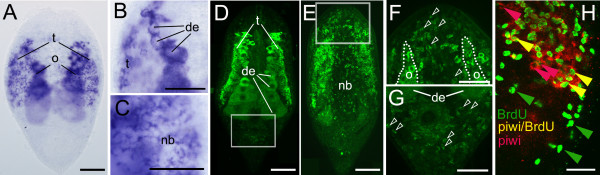
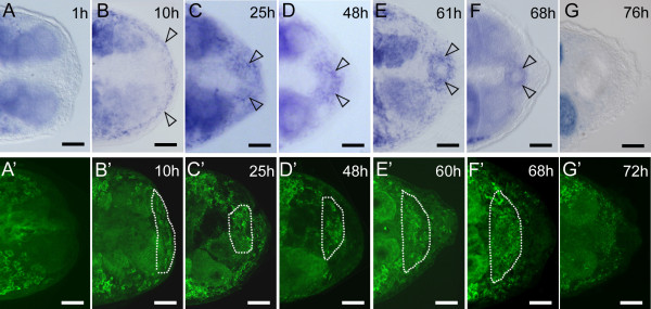
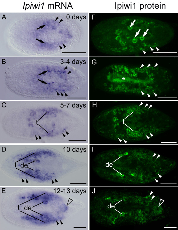
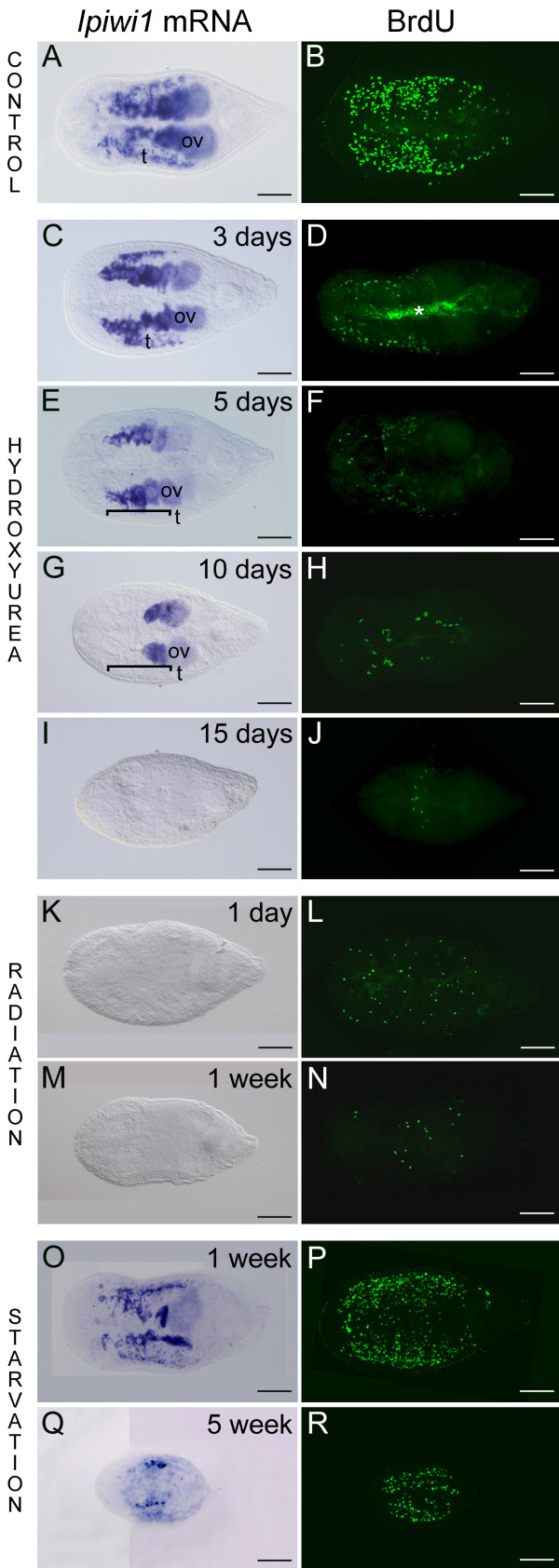
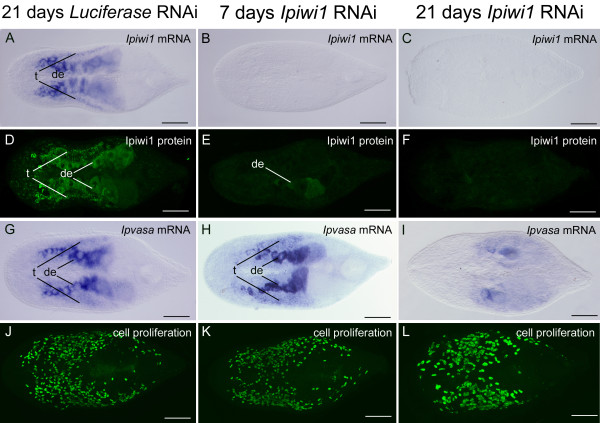
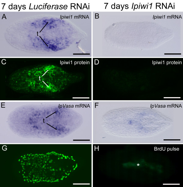
Results: We established enduring laboratory cultures of the acoel Isodiametra pulchra (Acoela, Acoelomorpha) and implemented in situ hybridization and RNA interference (RNAi) for this species. We used BrdU labelling, morphology, ultrastructure and molecular tools to illuminate the morphology, distribution and plasticity of acoel stem cells under different developmental conditions. We demonstrate that neoblasts are the only proliferating cells which are solely mesodermally located within the organism. By means of in situ hybridisation and protein localisation we could demonstrate that the piwi-like gene ipiwi1 is expressed in testes, ovaries as well as in a subpopulation of somatic stem cells. In addition, we show that germ cell progenitors are present in freshly hatched worms, suggesting an embryonic formation of the germline. We identified a potent stem cell system that is responsible for development, homeostasis, regeneration and regrowth upon starvation.

Conclusions: We introduce the acoel Isodiametra pulchra as potential new model organism, suitable to address developmental questions in this understudied phylum. We show that neoblasts in I. pulchra are crucial for tissue homeostasis, development and regeneration. Notably, epidermal cells were found to be renewed exclusively from parenchymally located stem cells, a situation known only from rhabditophoran flatworms so far. For further comparison, it will be important to analyse the stem cell systems of other key-positioned understudied taxa.


Figures

Figure 1
Figure 1
The stem cell system of Isodiametra pulchra (A, B). Morphology (C-E), distribution (F-I), and differentiation (J-L) of neoblasts. (A) Schematic drawing. (B) Differential interference contrast image. (C) Typical neoblast with nucleus (red) and thin rim of cytoplasm (yellow). (D-D") Macerated BrdU labelled cells show typical neoblast like morphology (E) BrdU labelled neoblast, as shown by immunogold staining after a 30 min BrdU pulse; arrowheads point to gold particles (F) histological cross section; brown spots are BrdU labelled S-phase cells. (G, H) Confocal projection overview (G) and detail of lateral body margin (H) after 30 min BrdU pulse; the red spot in (H) is a mitotic figure. Note that S-phase cells were lacking in the epidermis (between dotted lines). (I) Electron microscopic image of a posterio-lateral body margin. (J) Histological section, 10 days after the initial BrdU pulse. Some of the neoblasts underwent differentiation into epidermal cells (arrows); (K) BrdU labelled cells, differentiated after 10 days chasing time. Differentiating spermatid (top left), epidermal cells (top middle), parenchymal cell (top right), nerve cells (bottom left), and a muscle cell (bottom right) (L) BrdU labelled differentiated epidermal cell after 10 days chasing; arrowheads point to gold particles. bwm, body wall musculature; c, cilium; cc, condensed chromatin; cs, central syncytium; d, diatoms; e, egg; de, developing eggs; ep, epidermis; g, golgi; m mitochondria; mo, mouth opening; n nucleolus; st, statocyst; tw, terminal web. Scale bars (A, B, G) 100 μm; (C, E, L) 1 μm; (D, H; K) 10 μm; (F, J) 25 μm; (I) 5 μm.
Figure 2
Figure 2
Ipiwi1 mRNA expression (A-C) and protein localization (D-G) and BrdU/ipiwi1 (H) double labelling in I. pulchra. (A) Whole mount ipiwi1 in situ hybridization of an adult specimen. (B) Detail of developing eggs (de) and testes (t). (C) Dorsal focal plane showing ipiwi1 mRNA expressed in neoblasts (open arrowheads). (D, E) Confocal projections of Ipiwi1 protein localisation in testes and developing eggs (D) and in neoblasts (nb) (E). (F) Detail of the anterior region of (E) demonstrating Ipiwi1 positive cells (open arrowheads). (G) Detail of the posterior region of (D) demonstrating Ipiwi1 positive cells (open arrowheads). (H) Double staining of stem cells in S-phase (green) and ipiwi1 positive cells (red). Confocal projection (1,02 μm) shows the presence of BrdU-only labelled cells (green arrows), ipiwi1-only labelled cells (red arrows) as well as BrdU/piwi double labelled stem cells (yellow arrows). In all figures, anterior is to the top. Scale bars (A, D, E) 100 μm; (B, C, F-H) 50 μm.
Figure 3
Figure 3
Ipiwi1 mRNA expression (A-G) and protein localization (A'-G') during posterior regeneration. One hour after cutting (A, A'), ipiwi1 expression could not be detected at the regeneration site. After 10 hours, ipiwi1 was upregulated below the epidermis (arrows) (B, B'). At 25 hours postamputation (C, C') a significant proportion of cells within the regeneration blastema were ipiwi1 positive. From 48 hours onwards ipiwi1 expression and protein were present in the differentiating genital blastema (open arrows in D-F'). (G, G') After 76 h, ipiwi1 expression reached default levels. Scale bars 50 μm.
Figure 4
Figure 4
Ipiwi1 expression (A-E) and Ipiwi1 protein (F-J) during postembryonic development of I. pulchra. In freshly hatched animals, a subset of somatic neoblasts was visible as small piwi expressing cells (arrowheads) beside six to eight larger strongly stained primordial germ cells (that also express nanos, see text) (arrows in A, B, F). Until day seven neoblast number increased, PGCs multiplied and gave rise to testes and ovaries. At day seven testes and developing eggs could be observed (C, H). At days 10 and 12, a chain of developing eggs was present medially and testes were present along the lateral margin (D, E, I, J). Note the accumulation of ipiwi1 in the genital blastema (open arrowhead in E, J) which gives rise to the genital organs. A similar genital blastema was observed during regeneration (see Figure. 4). In all pictures, anterior is to the left. t, testes; de, developing eggs; (asterisk) autofluorescence of digested diatoms in the central syncytium. Scale bars 100 μm.
Figure 5
Figure 5
Ipiwi1 mRNA expression and cell proliferation (BrdU) in controls (A, B), during hydroxyurea treatment (C-J), after irradiation (K-N), and during starvation (O-R). (C) Upon three days HU treatment ipiwi1 mRNA could not be detected in neoblasts but was still present in testes (t) and ovaries (ov). (E) At five days of HU treatment ipiwi1 mRNA was present in all cells of the ovaries (ov) but not in testes (t, bracket indicates region of testes). (G) After 10 days ipiwi1 mRNA remained only in mature eggs; (t, testes; bracket indicates region of testes) (G). After 15 days no ipiwi1 mRNA could be detected (I) The number of S-phase cells strongly decreased during hydroxyurea treatment (D, F, H) and only single S-phase cells were found after 15 days (J). Irradiation (K-N) resulted in a complete elimination of ipiwi1 expression (K, M) and a strong reduction in the number of S-phase cells after one day (L) and one week (N) post irradiation. During starvation (O-R), ipiwi1 expression and protein localization were weakly reduced after one week (O, P). After five weeks of food deprivation, dramatic degrowth led to a strong reduction of Ipiwi1 mRNA and BrdU (Q, R). In all figures, anterior is to the left. t, testes; o, ovaries. Asterisk marks autofluorescence of diatoms within the gut. Scale bars 100 μm.
Figure 6
Figure 6
Influence of Ipiwi1 RNAi on adult I. pulchra at seven and 21 days of ipiwi1 dsRNA treatment. RNAi with luciferase control dsRNA did not show any effect on the level of i piwi1 expression (A), Ipiwi1 protein (D), ipvasa expression (G) or cell proliferation (J). Ipiwi1 mRNA was abrogated after seven days or 21 days of RNAi (B, C). Ipiwi1 protein was strongly reduced after seven days of ipiwi1 RNAi (E) and was completely eliminated after 21 days of ipiwi1 RNAi (F). Ipvasa expression was still prominent after seven days of ipiwi1 RNAi (H) and became significantly reduced after 21 days of ipiwi1 dsRNA treatment (I). Cell proliferation remained high up to 21 days of ipiwi1 dsRNA treatment (K, L). The specimen in figure 7L was processed for in situ hybridization before immunocytochemistry and therefore the nuclei appear larger. This protocol however did not alter cell number. In all figures, anterior is to the left. (t) testes; (de), developing eggs. Scale bars 100 μm.
Figure 7
Figure 7
Effect of ipiwi1 RNAi on development of I. pulchra after seven days of ipiwi1 dsRNA treatment. As a control, RNAi with luciferase dsRNA was performed which did not lead to any change in ipiwi1 or ipvasa mRNA expression (A, E), Ipiwi1 protein (C) or cell proliferation (G). After seven days of ipiwi1 RNAi treatment, ipiwi1 and ipvasa mRNA and protein were drastically reduced (B, D, F) and cell proliferation had completely stopped (H). All ipiwi1 knock-down juveniles died before eight days of postembryonic development. In all figures, anterior is to the left. Autofluorescence of diatoms is marked with an asterisk. (t) testes. Scale bars 100 μm.

References

    1. Agata K, Umesono Y. Brain regeneration from pluripotent stem cells in planarian. Philos Trans R Soc Lond B Biol Sci. 2008;363:2071–2078. doi: 10.1098/rstb.2008.2260. - DOI - PMC - PubMed
    1. Ladurner P, Egger B, De Mulder K, Pfister D, Kuales G, Salvenmoser W. In: stem cells: from hydra to man. 1. Bosch TC, editor. Vol. 1. Springer; 2008. The stem cell system of the basal flatworm Macrostomum lignano; pp. 75–94.
    1. Pfister D, De Mulder K, Hartenstein V, Kuales G, Borgonie G, Marx F. Flatworm stem cells and the germ line: developmental and evolutionary implications of macvasa expression in Macrostomum lignano. Dev Biol. 2008;319:146–159. doi: 10.1016/j.ydbio.2008.02.045. - DOI - PubMed
    1. Eisenhoffer GT, Kang H, Sanchez AA. Molecular analysis of stem cells and their descendants during cell turnover and regeneration in the planarian Schmidtea mediterranea. Cell Stem Cell. 2008;3:327–339. doi: 10.1016/j.stem.2008.07.002. - DOI - PMC - PubMed
    1. Handberg-Thorsager M, Fernandez E, Salo E. Stem cells and regeneration in planarians. Front Biosci. 2008;13:6374–6394. doi: 10.2741/3160. - DOI - PubMed

Publication types

Substances