. 2009 Dec 18;10(1):126.
doi: 10.1186/1465-9921-10-126.

Inhibiting toll-like receptor 4 signaling ameliorates pulmonary fibrosis during acute lung injury induced by lipopolysaccharide: an experimental study

Affiliations

Inhibiting toll-like receptor 4 signaling ameliorates pulmonary fibrosis during acute lung injury induced by lipopolysaccharide: an experimental study

ZhengYu He et al. Respir Res. .

Abstract

Background: Toll-like receptor 4 (TLR4) is essential in lipopolysaccharide (LPS)-induced fibroblast activation and collagen secretion in vitro. However, its effects on the process of lung fibroblast activation and fibrosis initiation during LPS induced acute lung injury (ALI) remain unknown. The goal of the present study was to determine the effect of inhibiting TLR4 on LPS-induced ALI and fibrosis in vivo.

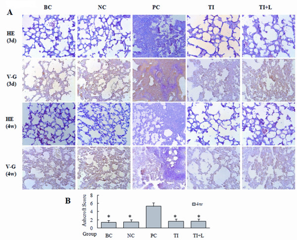
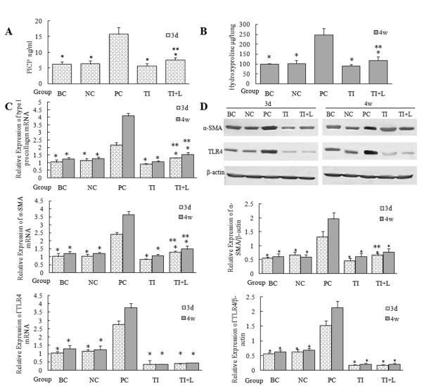
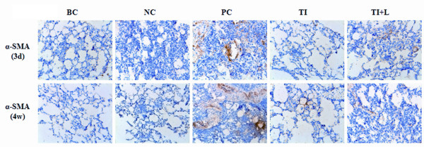
Methods: The ALI model was established by intraperitoneal injection of LPS in mice. TLR4-small hairpin RNA (shRNA) lentivirus was injected intravenously into the mice to inhibit TLR4 expression. mRNA and protein levels were detected by real-time PCR and Western-blot analysis, respectively. The contents of the C-terminal propeptide of type I procollagen (PICP) in bronchoalveolar lavage fluid (BALF) were detected by ELISA, and the degree of fibrosis was detected by van Gieson collagen staining, the hydroxyproline assay, and alpha smooth muscle actin (alpha-SMA) immunohistochemical staining.

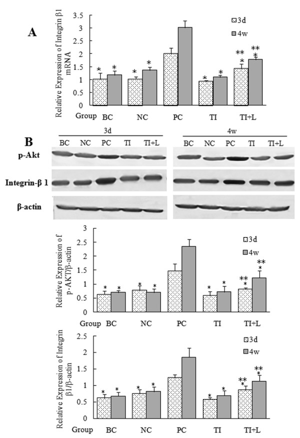
Results: Overexpression of TLR4, type I procollagen, alpha-SMA, and p-AKT in murine pulmonary tissue after intraperitoneal injection of LPS at 72 hours and 28 days were detected. Moreover, the degree of fibrosis was shown to increase by ELISA analysis of PICP in BALF, van Gieson collagen staining, the hydroxyproline assay, and alpha-SMA immunohistochemical staining. All of these changes were alleviated by intravenous infection with TLR4-shRNA lentivirus.

Conclusions: Inhibiting TLR4 signaling could ameliorate fibrosis at the early stage of ALI induced by LPS.


Figures

Figure 1
Figure 1
Inflammation and fibrosis in mouse lung tissue after LPS challenge. (A) The pathological changes in mouse lung tissue 72 hours and 28 days after intraperitoneal injection of LPS were observed by means of HE staining (HE, A, magnification ×200). Pulmonary fibrosis was observed by means of Van-Gieson staining (VG, magnification ×200). (B) The Ashcroft fibrosis score was used to compare the degrees of the pulmonary fibrosis 28 days after application of LPS in mice. The mice showed obvious inflammatory reactions in lung tissue 72 hours after LPS challenge. Typical pulmonary interstitial fibrosis appeared 4 weeks later. Infection with TLR4-shRNA lentivirus significantly inhibited the inflammatory reaction and fibrosis induced by LPS. BC: blank control group; NC: negative control group; PC: positive control group; TI: TLR4 inhibition group; TI+L: TLR4 inhibition group stimulated with LPS; 3d: specimens were collected 72 hours after LPS (or physiological saline) challenge. 4w: specimens were collected 28 days after LPS (or physiological saline) challenge. Each subgroup contains 6 specimens respectively (n = 6). Results were expressed as mean ± standard deviation indicated with column graph and error bar. Statistical significance was defined at p values < 0.05. *: P < 0.05 versus PC.
Figure 2
Figure 2
Collagen synthesis and secretion in mouse lung tissue after LPS challenge. (A) ELISA was used to detect PICP contents in mouse BALF to compare the synthesis of pulmonary type I collagen 72 hours after intraperitoneal injection of LPS. (B) Pulmonary collagen secretion and deposition 28 days after application of LPS in mice were quantitatively analyzed with hydroxyproline assay. (C) The mRNA expression of type I procollagen, α-SMA, and TLR4 in mouse lung tissue was detected by real-time PCR. (D) Protein expression of α-SMA and TLR4 were detected using Western blots. Type I procollagen, α-SMA, and TLR4 expression in mouse lung tissue increased significantly 72 hours after LPS challenge and become intensive 4 weeks later. Infection with TLR4-shRNA lentivirus significantly inhibited these changes. BC: blank control group; NC: negative control group; PC: positive control group; TI: TLR4 inhibition group; TI+L: TLR4 inhibition group stimulated with LPS; 3d: specimens were collected 72 hours after LPS (or physiological saline) challenge. 4w: specimens were collected 28 days after LPS (or physiological saline) challenge. Each subgroup contains 6 specimens respectively (n = 6). Results were expressed as mean ± standard deviation indicated with column graph and error bar. Blots were representative of six separate experiments. Statistical significance was defined at p values < 0.05. *: P < 0.05 versus PC; **:P < 0.05 versus TI.
Figure 3
Figure 3
Expression of α-SMA in mouse lung tissue after LPS challenge. Paraffin-embedded mouse lung tissue samples were deparaffinized, rehydrated, and processed to detect α-SMA using the avidin-biotin immunoperoxidase method. Representative images (×200) of samples are shown. BC: blank control group; NC: negative control group; PC: positive control group; TI: TLR4 inhibition group; TI+L: TLR4 inhibition group stimulated with LPS; 3d: specimens were collected 72 hours after LPS (or physiological saline) challenge. 4w: specimens were collected 28 days after LPS (or physiological saline) challenge. Each subgroup contains 6 specimens respectively (n = 6).
Figure 4
Figure 4
Activation of the PI3K-Akt pathway and expression of integrin β1 in mouse lung tissue after LPS challenge. The p-AKT or integrin β1 in mouse lung tissue was detected by real-time PCR (A) or Western blot (B). P-Akt, which reflected Akt phosphorylation levels, and integrin β1 expression increased significantly 72 hours after LPS challenge and become intensive 4 weeks later. Infection with TLR4-shRNA lentivirus significantly inhibited these changes. BC: blank control group; NC: negative control group; PC: positive control group; TI: TLR4 inhibition group; TI+L: TLR4 inhibition group stimulated with LPS; 3d: specimens were collected 72 hours after LPS (or physiological saline) challenge. 4w: specimens were collected 28 days after LPS (or physiological saline) challenge. Each subgroup contains 6 specimens respectively (n = 6). Results were expressed as mean ± standard deviation indicated with column graph and error bar. Blots were representative of six separate experiments. Statistical significance was defined at p values < 0.05. *: P < 0.05 versus PC; **:P < 0.05 versus TI.

Similar articles

Cited by

References

    1. Marshall RP, Bellingan G, Webb S, Puddicombe A, Goldsack N, McAnulty RJ, Laurent GJ. Fibroproliferation occurs early in the acute respiratory distress syndrome and impacts on outcome. American journal of respiratory and critical care medicine. 2000;162(5):1783–1788. - PubMed
    1. Janardhan KS, McIsaac M, Fowlie J, Shrivastav A, Caldwell S, Sharma RK, Singh B. Toll like receptor-4 expression in lipopolysaccharide induced lung inflammation. Histology and histopathology. 2006;21(7):687–696. - PubMed
    1. Yoshizaki A, Iwata Y, Komura K, Ogawa F, Hara T, Muroi E, Takenaka M, Shimizu K, Hasegawa M, Fujimoto M. et al.CD19 regulates skin and lung fibrosis via Toll-like receptor signaling in a model of bleomycin-induced scleroderma. The American journal of pathology. 2008;172(6):1650–1663. doi: 10.2353/ajpath.2008.071049. - DOI - PMC - PubMed
    1. Armstrong L, Thickett DR, Mansell JP, Ionescu M, Hoyle E, Billinghurst RC, Poole AR, Millar AB. Changes in collagen turnover in early acute respiratory distress syndrome. American journal of respiratory and critical care medicine. 1999;160(6):1910–1915. - PubMed
    1. Olman MA, White KE, Ware LB, Simmons WL, Benveniste EN, Zhu S, Pugin J, Matthay MA. Pulmonary edema fluid from patients with early lung injury stimulates fibroblast proliferation through IL-1 beta-induced IL-6 expression. J Immunol. 2004;172(4):2668–2677. - PubMed

Publication types